Thursday 30 December 2021

Micro-worlds

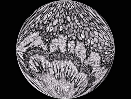
Sometimes it is nice to do something different with the images you observe through the microscope. Like in this case with a cut through the petiole of the Castanea sativa (chestnut).

Two photos were taken with the Motic BA410E. 

One with the Moticam S12, a 10X PlanApo objective, a polarization and a lambda filter. 

Another with the Moticam 285C Pro, a black and white camera. This camera is mainly intended for the recording of fluorescence images, under low light conditions. The rest is then a matter for the photo editing program.


The images are vaguely reminiscent of Mother Earth when viewed from space. You could call the images micro-worlds, so to speak. 

It is a pleasure to work with microscopy in such a way every now and then.

Friday 24 December 2021

Not just limestone

Certain sedimentary rocks such as limestone can be impregnated with hydrocarbons and can contain 4 to 9% bitumen or "shale oil". Asphaltic or bituminous limestone is therefore a mineral impregnated with natural bitumen. This mineral is mined from underground mines or via open-pit mining. 

From asphaltic limestone, bituminous oil or asphalt can be extracted for use in the cosmetic or pharmaceutical industry. This tough and sticky product is also mixed with other raw materials to produce the well-known black pavement for our roads. In addition, it is used for waterproofing of roofs, walls, terraces, sidewalks, bridges, casings, tanks, etc. It is also a good acoustic insulator that is able to absorb the noise of machines.

Apart from the mentioned applications, the bituminous oil was once considered a universal remedy, especially good for the treatment of colds, hair loss (Hahn oil), stomach diseases and last but not least, rheumatism. By distillation of asphaltic limestone, it is still possible to obtain gas and petroleum.

On the photo taken of a thin section we see Ooids with bituminous material (containing some fossils) around it. Ooids are spherical or subspherical carbonate grains characterized by an internal concentric structure. Long time ago, the layers or coatings were formed in agitated waters as these grains were rolling around, after which they were piled in layers, under pressure, to form sediment rock.


© www.willemsmicroscope.com

Thursday 16 December 2021

Caught and eaten by Portuguese Sundew

Portuguese Sundew or Drosophyllum lusitanicum is a shrub-like carnivorous plant that can grow up to 1.5 m in height, but is usually shorter (about 40 cm). The genus name, Drosophyllum, is derived from the Greek "drosos" (meaning dew) and "phyllon" (meaning tribe or leaf). This name is a reference to the glittering droplets on the catch stalks. 

The species name is derived from Lusitania, the ancient name for Portugal. In its native habitat, Drosophyllum is relatively rare, with plants growing in narrow coastal or maritime regions (to maximum a few tenths of kilometers from the coast) with regular morning fog during summer. 

Besides Portugal, Drosophyllum can be found in Andalusia and in northern Morocco.

Portuguese Sundew is a very interesting and curious plant for several reasons. It has two types of glands on the leaves; the huge stalked glands - as shown on the image - that attract and trap prey and the sessile, digestive glands. 

Compared with other carnivorous plants, it is one of the most successful ‘hunters’ in terms of quantity of captured prey, and all with the absolutely passive primitive adhesive traps (the stalked sticky glands). 

Production of the mucilage for trapping prey is so intense that it often drips from the leaves. Digestion of prey is very rapid, being complete within several days. Luring is accomplished by the sweet, honey-like odor of the mucilage, detectable by the human nose.


© www.willemsmicroscope.com

Wednesday 15 December 2021

New minerals for a classic mine in Occitània: Correc d’en Llinassos, Oms, France

Oms is a beautiful village, in the ancient region of Roselló (nowadays Eastern-Pyrenees, France), in the south of historic region of Occitània. This locality includes different mining sites of scientific interest. One of these mining operations is in the Correc d'en Llinassos (torrent), near Oms (mindat.org, loc-49065).

This deposit was studied by the AFM (Association Française de Microminéralogie) in the middle of the last decade (Berbain & Favreau, 2007). In this article, numerous species were citated, the most interesting containing minerals were the Ni: annabergite, bottinoite, gersdorffite, glaucospherite, millerite, ullmannite. It is necessary to add one more that was identified for the first time worldwide: omsite, named after this locality (type locality) (Mills et al., 2017). This is a very rare hydroxyantimonite of iron, nickel and copper, member of the cualstibite group. Correc d’en Llinassos is also the type locality for hydroxyferroroméite, identified in 2017 (Mills et al., 2017a).

During a visit to the mine, with prior authorization from the owner of the farm, since access is prohibited, it was possible to collect various mineral specimens from the outer area of the mine, to later be studied. Among them, some globular aggregates of white color on siderite stood out, which after being analyzed by SEM-EDS indicated that it could be some magnesium carbonate. The Raman spectra obtained were consistent with dypingite or hydromagnesite. To close the study, a powder X-ray diffraction was carried out. Both the spectrum and the cell yielded values confirmed that it was hydromagnesite. This mineral had not been described in this mine.   

Globular white aggregates of hydromagnesite accompanied by Mg rich malachite

XRD spectrum (powder): hydromagnesite from Oms. 
Courtesy: Geociències Barcelona, Geo3BCN–CSIC, Bruker D8-A25 (Cu Kα, PSD detector)

On the other hand, it was possible to observe several globular aggregates of green colour that could correspond a visu with malachite. Reaction with HCl indicated that it was a carbonate. A copper-nickel hydroxylcarbonate from the Rosasite group has been described in this deposit, which could be identified as glaucospherite, by appearance and chemical reaction. Raman spectra also indicated that it was a similar mineral malachite. The studies were carried out using SEM-EDS and the results were very interesting since, surprisingly, most of these malachite aggregates contain a certain percentage of magnesium (Mg: Cu 1: 4-6).

Green globular aggregates of magnesium rich malachite with aragonite (white)


SEM-EDS uncoated. Malachite Mg-rich 
Cortesy: Geomar-Enginyeria del Terreny, SEM-EDS Phenom G5 XL

Its resemblance to the photographs published as glaucospherite from this deposit suggests that, in some cases, without chemical analysis it may be difficult to identify one or the other mineral. After reviewing numerous specimens, it was possible to find globular aggregates of a sky blue to bluish green color, in which magnesium and copper were found in 1: 1 proportion. It was, unsurprisingly, identified as McGinnessite.

Globular aggregates of blue-green mcguinnessite acompanied by Mg-rich malachite (green)

SEM-EDS uncoated. Mcguinnessite from Oms. 
Cortesy: Geomar-Enginyeria del Terreny, SEM-EDS Phenom G5 XL

Mineral deposits, although studies have been published on them, do not cease to amaze. Continued study will enrich the country's mineralogical heritage and contribute a grain of sand to science. Obviously, also supporting the collaboration between academic mineralogists and amateur mineralogists, mineralogists after all, is our duty and obligation. 


Acknowledgments
Thanks to Geomar-Enginyeria del Terreny for SEM-EDS studies; Dr. Jordi Ibáñez and Soledad Álvarez, Geociències Barcelona (GEO3BCN - CSIC); Dr. Tariq Jawhari, Raman departament of Centres Científics i Tecnològics de la Universitat de Barcelona (CCiTUB); Dr. Joan Carles Melgarejo (UB) to facilitate the study of the samples and to the owner of the property where the mine is located.


Bibliographical notes
- Berbain, C., Favreau, G. (2007): “Un exemple peu courant de minéralisation nickélifère: le Correc d'en Llinassos à Oms (Pyrénées-Orientales)”. Le Cahier des Micromonteurs, 95, 1, 3-24.
- Mills, S.J., Kampf, A.R., Housley, R.M., Favreau, G., Pasero, M., Biagioni, C., Merlino, S., Berbain, C., Orlandi, P. (2012): “Omsite, (Ni,Cu)2Fe3+(OH)6[Sb(OH)6], a new member of the cualstibite group from Oms, France”. Mineralogical Magazine, 76, 1347-1354.
- Mills, S.J., Christy, A.G., Rumsey, M.S., Spratt, J., Bittarello, E., Favreau, G., Ciriotti, M.E., Berbain, C. (2017a): “Hydroxyferroroméite, a new secondary weathering mineral from Oms, France”. European Journal of Mineralogy, 29, 307-314.
- Mindat.org: “Correc d'en Llinassos (Ravin d'en Llinassous), Oms, Céret, Pyrénées-Orientales, Occitanie, France”. https://www.mindat.org/loc-49065.html [on line 11/2021].

Thursday 9 December 2021

Phacus longicauda

Phacus longicauda is a species of flattened, pointed single-celled protist microalgae belonging to the phylum Euglenozoa. It is a solitary and free-swimming algae.

Phacus longicauda has a very striking and rigid periplasm, with spiral bands, and numerous small disc-shaped chloroplasts. The cell is flat and rigid and powered by a flagellum at the front of the cell. There is a highly visible red eye spot near the flagellar base. These red eyespots are present in most Euglenoïds.

Phacus longicauda is found in freshwater habitats around the world. These habitats include ponds, pools and standing water. An organically enriched freshwater environment is essential for the development of these species.

Phacus species sometimes have a peculiar twisted shape. They are photosynthetic unicellular organisms, which means that they are able to produce their own food. Although the genus receives their nutrients primarily through photosynthesis, they are also able to feed on certain types of algae and bacteria.

Most of these organisms also have a semi-rectangular eyespot, often reddish in color, and a single flagellum, although some species have two that emerge. The flagellum is responsible for movement of the cell by rotating in the direction of travel, which allows the cell to slide and swim in the water.

At the base of the flagellum, adjacent to the eyespot, lies the paraxial swelling that functions in photoreception. It is the intricate coordinated interactions of the eyespot, paraxial swelling and some other parts of the cell that enable the cell to swim to and from the light. The ‘eyespot’ designation is therefore somewhat misplaced because it is only part of the photosensitive device and is not the photoreceptor itself, which is the paraxial swelling.

For detailed descriptions, see the standard work "Freshwater Algae of North America, Ecology and Classification".


Friday 3 December 2021

How does it look after 4 years at about 1200 ºC?

The pictures taken with the SMZ-171 and the Moticam 10+, are showing a Haynes alloy component which has been exposed to a temperature of about 1200 oC during a period of 4 years. The component was positioned in a naphtha cracking oven (methane fuel side) of a chemical ethylene plant. On the surface of the component that is affected greatly, carburization and crystal formation can be seen. By means of ultrasonic thickness measurement it can be checked if sufficient unaffected material is left under the carburized layer, in order to see if the component is still strong enough.


Haynes 230 alloy is a nickel-chromium-tungsten-molybdenum alloy that combines excellent high-temperature strength, outstanding resistance to oxidizing environments up to 2100°F (1149°C) for prolonged exposures, premier resistance to nitriding environments, and excellent long-term thermal stability. It is readily fabricated and formed, and is castable. Other attractive features include lower thermal expansion characteristics than most high-temperature alloys, and a pronounced resistance to grain coarsening with prolonged exposure to high temperatures.

Rod material out of Haynes alloy

Exposed Haynes alloy piping distance piece

Haynes alloy 230 is composed of  Ni 57%, Cr 22%, Tu 14% and Fe < 3%

With thanks to: Gerard Janssen and Rijk Koster, mechanical engineers

© www.willemsmicroscope.com

Monday 29 November 2021

A Motic tablet camera seen from a different perspective

If you would like to take a closer look at the screen of your tablet - in this case it is a Motic tablet-camera combination - then you could very well use a microscope. 

The screen of the tablet was observed through the Motic stereomicroscope SMZ171 for the moving images. The Motic BA410E was used with a LM Plan 10X objective for the still images.

The camera used to record the color switching of the pixels was the Moticam 1080 and the still images were taken with a Moticam 10 camera. You can see the typical pixel shape, which is different from the one you can see on e.g. mobile phones. Red, green and blue are the basic colors used. Each pixel can switch to only one of these colors, but combinations thereof will form a complete color spectrum.


Thursday 25 November 2021

Pigment protects

The skin is the largest organ of the human body. The skin consists of three layers. The top layer is formed by the epidermis. The middle layer is called the dermis. The bottom layer is the subcutaneous connective tissue. The epidermis consists mainly of special skin cells called the keratinocytes. The keratinocytes are formed in the lower layer (the basal layer) and slowly slide upwards from there. Gradually they transfer to a dead keratinized cell layer, the horny layer, where the cells lie loosely against each other.


Melanocytes are present in the epidermis, in addition to the keratinocytes. Melanocytes are pigment cells that lie between the cells of the basal cell layer and make the pigment grains that are transferred via offshoots to the keratinocytes. The pigment of the pigment granules, the melanin, determines to a large extent the color of the skin and protects us from sunlight. The more pigment grains, the darker the skin. Pigment is a brown dye that is produced by the pigment cells in the lower layer of the epidermis. Pigment cells lay therefore relatively superficial in the skin. In the pigment cell, the pigment is packaged in pigment granules, and thus delivered to the upper epidermal cells. Only when the pigment is in the epidermal cells the skin is visibly pigmented.

The functions of pigment cells are besides determining the skin color also protecting the skin against harmful ultraviolet (UV) rays. Under the influence of UV radiation, the number of active pigment cells and pigment production increases. The result is a brown discoloration of the skin.

With thanks to Dr. Eddie Wisse and Frank van Campen, members of the Royal Antwerp Society for Micrography.
Prepared slide by Lieder www.lieder.com

© www.willemsmicroscope.com


Tuesday 23 November 2021

Snails: Powerhouses

Snails are the only mollusks that can live on land. As many as 60,000 to 75,000 species are known in the world. Snails are also called gastropods. This is because the muscles on the abdomen (bottom of the snail) are responsible for the movement. Most snails are hermaphrodite, which means that they have both male and female sexual organs. Some snails are even solitary and can reproduce without needing another snail!


Believe it or not, but a snail has more teeth than a shark! A snail can have more than 25,000 teeth and sharks "only" have 3,000. The snail hides all these small teeth on its tongue (radula)_ and scrapes small pieces off the leaves that it eats. Ever wondered why there are so many holes in the leaves of your plants? A snail can eat up to half its own body weight in one day! It does not matter much what snails eat. They prefer to eat only the soft part of the plant, but when there is a long drought, they can take a bite from your newspaper if they can't find anything else. They are not picky.


© www.willemsmicroscope.com

Thursday 11 November 2021

Artery from another perspective

The preparation of the artery shown in both images is of unknown origin and the staining method is unknown as well.

A fluorescence image of an artery tissue as shown here is not usual. The preparation is more or less evenly green fluorescent colored, because the dye used has attached itself to the structural parts of the preparation. We see a fluorescence color image with details as you might encounter in bright field microscopy with dyes such as e.g. toluidine blue. Such dyes show more or less the same details in the preparation with ordinary bright field microscopy as is shown in the first image.

As mentioned before, visualizing a histological specimen completely by using a fluorescent substance is unusual, because here you apply only one color that shows no specific adhesion, except for proteins, because e.g. fat is uncolored in this preparation. With a normal hematoxylin and eosin staining you have more differentiation, namely in the cell core (DNA, dark blue) and proteins, cytoplasm (pink)


In histology, fluorescence is mainly used after a fluorescent substance has been chemically attached to a specific component present in the tissue.

On the fluorescent image of the preparation the parts of an artery can also be seen. The lumen is filled with green colored red blood cells, then there is a very thin (barely visible) layer of endothelium, the meandering lamina elastica interna (clearly visible) a layer of smooth muscle, the thinner lamina elastica externa (clearly visible), the adventitia (connective  tissue) that turns into adipose (fat) tissue. 


© www.willemsmicroscope.com


Thursday 21 October 2021

Thousands of small lenses work together

Seen from the front, the head of the honeybee has a triangular shape, the head of the drone it is more round. On the head are the eyes, antennae and mouth parts. Important glands are located in the head and the main center of the nervous system: a nerve bud that serves as a brain.


Friday 15 October 2021

A switching station

Ganglia are part of the peripheral nervous system. The peripheral nervous system includes all nerves outside the central nervous system (bundles of long nerve shoots with supporting cells) and small centers of nerve cells the ganglia.

A ganglion is a combination of mainly cell bodies (ganglion cells) with the nucleus of the nerve cell surrounded by cytoplasm without further shoots. This cell body with the nucleus as its center forms the metabolic center of the cell and is sensitive to stimuli. Ganglia function as switching stations for nerve impulses.

Wednesday 6 October 2021

A pest from Africa

The southern green stinkbug (Nezara viridula) is a relatively new pest that originates from Ethiopia. The pest has a large number of host plants, but in horticulture this bug is mainly found in bell pepper cultivation. In the first instance, in The Netherlands, the southern green stink bug was only spotted at the end of the summer, but the bug has also shown itself earlier in the season in recent years.


Females lay around 30 to 130 eggs at a time. They do this in "glued together", yellow and white clusters at the bottom of the leaf. If they are just laid, they look like little pearls. Depending on the temperature, the eggs hatch after 4 days to 3 weeks and the empty eggs remain on the leaf. The nymphs initially stay together and do not yet affect the plant. They then go through different stages, each time shedding their skin, with each stage having a different appearance. In the fifth stage, the bugs begin to spread over the plant and eat it.

Wednesday 29 September 2021

Ficus carica petiole and Wacker 3A staining

Vascular bundles can be seen very well in these cross-sectional views of a leaf stem of the fig tree, thanks to the Wacker 3A triple staining. This coloring is done with a mixture containing Astra blue, Acriflavin and Acridine red.


We see the vascular bundles that lie in an outer circle, characteristic of (eu)dicotyledonous plants. In monocotyledonous plants, the bundles are irregularly distributed over the cross section. The Xylem vessels lie on the inward side and the Phloem vessels on the outside of the outer circle. Between Xylem and Phloem we see the intravascular Cambium. Vascular bundles are also present in the inner basic Parenchyma.

Due to the good uniform thickness of the coupe and the fine quality of the Moticam S12 camera, the photos could be taken without stacking.

Tuesday 21 September 2021

Sundew

Sundew or Drosera forms perfect rosettes to the ground and has red tentacles with a sticky, glittering droplet which is secreted by a gland. The flower owes its name to this. Small animals get stuck in the drop and are pushed by the moving tentacles to the leaf surface where they are digested.


Prepared slide by Lieder

© www.willemsmicroscope.com

Monday 13 September 2021

Kahler's disease

The disease owes its name to the Austrian doctor Otto Kahler, who described the disease as one of the first. Kahler's disease, also called multiple myeloma, is a disease of the bone marrow caused by an uncontrolled proliferation of a certain type of white blood cells: plasma cells (also called plasmocytes). Plasma cells are responsible for the formation of antibodies under normal conditions.

The bone marrow is a spongy substance located in the interior of bones, especially the pelvis, sternum, ribs and vertebrae. Bone marrow plays a role in forming bones, but also in forming the cells of the blood: white blood cells (leucocytes), red blood cells (erythrocytes) and platelets (thrombocytes). The disease develops in one abnormal plasma cell, which divides uncontrollably and whose offspring also divides uncontrollably. Since they are all related, they only synthesize one specific type of antibody (or part of it). Since antibodies are proteins, the antibody produced is referred to as the "M protein" (from Myeloma protein). The name paraprotein is also commonly used for the M protein. When a specific piece (called “light chain”) of a paraprotein is found in the urine (or blood serum), it is called the Bence-Jones protein.

Friday 10 September 2021

It grows on tree trunks

Dilated scalewort or Frullania dilatata grows mainly as a pioneer on trees with a moderately nutrient-rich bark. The species is somewhat drought-tolerant and can be found on free-standing trees, except in forests. Especially in full sun, the plants have their characteristic rust brown color. This moss experienced a decline in the last century due to air pollution, but is now common in almost all parts of The Netherlands. It is an attractive species, which is not easily overlooked in inventories. As a result, the distribution picture does suggest a greater number of occurrences than is actually the case: regularly there is only a single find per square section of land of 1 x 1 km.


© www.willemsmicroscope.com

Friday 3 September 2021

Tempskya, growing millions of years ago

Tempskya is an extinct genus of tree-shaped ferns, the fossils of which have so far only been found in Cretaceous layers. The Cretaceous is a geologic period and system that spans 79 million years from the end of the Jurassic period 145 million years ago to the beginning of the Paleogene period 66 million years ago.


The stem-shaped structure of Tempskya is called "false stem" because it is formed by an entanglement of large numbers of stems and roots. The upward growing stems, which have a diameter of 2.5-10 mm, gave large numbers of roots (diameter about 1 mm) that grew downwards. They formed a felt-like mass around the stems. Because the stems branched regularly, a very solid whole was created. On a cross section of a trunk (see the images) the relatively large stems and the small roots can be seen.


Thursday 26 August 2021

Which freshwater algae do we see?

In the video you will find some algae found in fens in the ‘Wortel Kolonie’ located in Belgium. The ‘Wortel Kolonie’ is a beautiful protected nature reserve with a complex of buildings that served as an asylum for vagrants long time ago. (Here you will find the information in Dutch, English, French and German).

This nature reserve contains a few shallow fens. On a summer day in July, some water samples were taken from two fens using a trawl net with a mesh size of 30 microns. Without examining these samples in depth, more than fifty different organisms, phyto- and zooplankton, were found. Only a few of them are shown in the video. In one of the algae we see a oogonium.

The names of the organisms in the video are deliberately omitted here, to give hydrobiology enthusiasts the opportunity to identify the organisms themselves.

Friday 20 August 2021

Drill a hole before you can eat

The whelk or Buccinum undatum (Linnaeus, 1758) belongs to the snails (Gastropoda).

The whelk is a large, thick predatory snail with 7 or 8 turns. A deep seam runs between the turns. The mouth opening takes up almost half of the shell and ends in a sipho. On the surface there are horizontal ribs and clear vertical growth lines. Often there are also vertical ribs at the top of the shell. It grows up to 11 cm high and 7 cm wide. The whelk has yellowish-white with dark spots. Whelks washed ashore are often dark blue, because they have been lying on the bottom of the sea for a long time.


Tuesday 17 August 2021

Vorticella - The fastest bell in the world?

Vorticella is a genus of single celled organisms living in both marine- and freshwater habitats. The members of the vorticella genus are commonly referred to as bell animalcules because of their shape. The thing that makes bell animalcules unique are their long stalks which they use to attach themselves to a substrate, which could be things like rocks, twigs and even small animals. The stalks of vorticella resemble our muscle fibers but are able to contract a lot faster than our muscles are. If the cell gets disturbed, it’s able to contract the stalk with an impressive speed of up to 6 meters per second as a defence mechanism to get away from danger or possibly scare predators away.

Thursday 12 August 2021

Built for speed

The peregrine falcon is one of the fastest birds in the sky. During level flight, with motion generated by wing-beats alone, they can reach speeds between 60-100 km/h. This is one of the fastest known speeds for level flight.

Tuesday 3 August 2021

Mint

Mentha spicata (Green Mint) is a low, creeping and rather invasive plant that is indispensable in a fragrant border. Mentha spicata blooms with small pale purple or pink flowers. It easily grows widely on all soil types and will certainly have to be kept under control. It is an herb with numerous uses, both kitchen use and medicinal. On the photo you can see covering hairs and glandular trichomes from which the typical aromatic substances are excreted.


© www.willemsmicroscope.com

Tuesday 27 July 2021

One Coleps makes two, cell division

  • Alga: Coleps
  • Order: Prorodontida Corliss, 1974
  • Family:  Colepidae Ehrenberg, 1838
  • Genus: Coleps Nitzsch, 1827


Coleps is a barrel-shaped ciliate, notable for its regularly arranged ectoplasmic shields, or platelets, of calcium carbonate. The covering with the scales is sometimes completely or partly absent immediately after an amitotic division (direct cleavage of the nucleus without the formation of mitoses). This is clearly visible in the accompanying video. Cell division took approximately 45 minutes. The color of the cell is usually brown and is less determined by the color of the food consumed than with other ciliates.

Wednesday 21 July 2021

Giant chromosomes

Giant chromosomes (or polytene chromosomes) are found in the cells of larvae of flies and mosquitoes, in springtails and in some other invertebrates. In mosquitoes we find them, for example, in the cells of the bug Chironomus and in the cells of flies like Drosophila (fruit fly). The growth of these larvae occurs because the cells grow in size, not in number. The chromosomes also grow in size because the DNA multiplies many times without the cell or nucleus dividing. A giant chromosome therefore consists of hundreds to thousands of undivided chromatides. The chromatids have a sequence of light and dark bands, the so-called chromomers. Due to the dimensions of the giant chromosomes, these bands are very noticeable. On the basis of these bands it is easy to indicate where the genes are located on the giant chromosome (called ‘gene mapping’)


On the image, swellings, so called puffs, can be seen at particular sites along the length of the polytene chromosome. These are diffuse uncoiled regions where transcription is actively taking place.

Polytene = composed of many chromatides
Chromatides = the two identical parts of a chromosome
Gene = a piece of DNA containing certain hereditary characteristics
© www.willemsmicroscope.com

Thursday 15 July 2021

New Zealand flax

Phormium tenax or New Zaeland flax are evergreen perennials, making a large clump of leathery, strap-shaped leaves coming to a decided point, with tall panicles of small, tubular flowers in summer.

New Zealand flax is an excellent fiber plant, hence the association with real flax (Linum usitatissimum) that is grown in Europe. The fibers of the leaves, up to 2 m long, were used by the Maori to make clothes, bags and shoes, and later the Europeans made rope and sails. All parts of the plant were used: for medicine made from roots, face powder from flower pollen and old blooming stems roped together as rafts.


Wednesday 14 July 2021

What exactly is the function of our red blood cells?

Red blood cells (RBCs) also known as erythrocytes are the most abundant cell in the human body. With a staggering number of 25 trillion cells, yes TRILLION, the red blood cells make up around 80% of all your cells. The reason why we have so many of them is because they have a few incredibly important functions critical to our life.

Notice the characteristic donut-shape.

Friday 9 July 2021

Cornwallite, conicalcite and other minerals from the Reconquistada prospect, Dolores mine, Pastrana, Mazarrón, Murcia

The “Reconquistada” claim is made up of small extractive mining works near the “Dolores” iron mine, in the municipality of Pastrana (Murcia, Spain). In this area there are various exploitations that benefited copper ores. In the case of the “Reconquistada” mineral species, the presence of arsenic in the mineral composition made copper extraction difficult.

From a scientific point of view, these mining works have been important due to the numerous species found. Three of them were identified for the first time worldwide in this mines: barahonaite-(Fe), barahonaite-(Al) and cobaltarthurite (Jambor et al., 2002; Viñals et al., 2008).

But there are other interesting and very colourful minerals. Two of them are very similar in appearance, but distinguishable under magnification: cornwallite and conichalcite. Both are copper arsenates, but conichalcite contains calcium. Cornwallite appears as spherical globules, with a smooth and very brilliant surface, usually with a lighter green color than conichalcite. Conichalcite tends to have a rough surface and a more intense colour.

In this specimen of the photograph, various botryoidal aggregates of conichalcite could be observed, with the characteristics described before, together with an isolated sphere of cornwallite. They are disposed on a matrix with yukonite (an iron and calcium arsenate).

Globular aggregates of conichalcite with internal radiated crystals

Monday 5 July 2021

Spicules bring light

Sponges, like all animals, possess some sort of a skeleton that gives their bodies shape. As a whole, poriferans have diverse skeletal elements including calcareous laminae, organic filaments, and siliceous and calcareous spicules. The skeletons of each of the major poriferan groups are distinctive and have been used to reconstruct their evolutionary relationships.

This is a second brief publication about spicules, but this image is completely different however; here the darkfield technique has been adapted, using a turret condenser.

Wednesday 23 June 2021

Stamens and a jewel clip

If anything stands out about the bunch of lilies pictured below, it is the large dark coloured stamens. These are worth seeing under the stereo microscope. There is a handy tool to enable this. 


Thursday 17 June 2021

The Rose of Jericho ‘a plant full of fire’

The Dutch Society for Microscopy (NGVM) department West, organized a workshop on fluorescence microscopy. An introductory presentation was given on this important application of microscopy. Fluorescence microscopy is used in many different fields, such as in medical science, for example in cytology, but also in other areas such as forensic research, pollen research, mineralogy, etc.

Wednesday 9 June 2021

Daphnia’s maneuvers in the dark

Like many other crustaceans, water fleas are very sensitive to light. They have a light-sensitive organ, also called Ocellus, with which they can swim in the direction of the light.

Friday 4 June 2021

The yak keeps you warm and tidy in the winter

The yak is a bovine species, that lives amongst others in Tibet. They graze in the cool higher part of mountainous areas. Yaks are not adapted to living in hot climates. The main raw material produced by the yaks is wool, existing of tail hair and long hair along the yak belly. Yak shearing takes place in the spring, which is a difficult action because of the furious personality of this animal. 

Wednesday 26 May 2021

Chaoborus larva

These are free-swimming larvae of so called phantom midges or lake flies and inhabit the area between the bottom and the surface of ditches. They are hunters of small crustaceans such as daphnia and cyclops. These animals are practically transparent, and are therefore also called glass larvae. (For the sake of clarity, the preparation has been stained)

With the help of air bubbles in the fore and abdomen, they remain in balance with the water and float unseen through ditches, swimming with leaping movements. They grow to about 1 cm to 1.5 cm long. When two larvae meet each other, a tumble around each other occurs.

Wednesday 19 May 2021

Multipurpose symbiosis

The black alder - Alnus glutinosa - lives in symbiosis with bacteria from the actinomycetes group. These are capable of withdrawing nitrogen from the atmosphere. Through this partnership, the alder, despite its relatively high nutritional requirements, can colonize nitrogen-poor locations.


Monday 17 May 2021

Algae Bloom


These beautiful golden spheres are single colonies of the golden algae synura. Like all other organisms, synura is able to reproduce and one colony eventually turns into two. But when conditions are right, reproduction happens a lot more frequently and what used to be a few colonies can quickly turn into an enormous algae bloom. Even though each synura colony is microscopic and only measures around 20-30 microns in diameter. Their large numbers in an algae bloom are able to turn the water in their freshwater habitats a yellowish brown color. A bloom of synura like the one in the image usually happens in spring and fall when the water is cold. But not all algae will bloom in cold water.