
Wednesday, 28 December 2016
‘Flowers’ beneath the surface
Tuesday, 13 December 2016
No watch without a microscope

Friday, 2 December 2016
A colon, why?
 |
| Human colon | SMZ171 stereomicroscope | Moticam 10 |
In the large intestine (colon) resorption takes place of the remainders of the digested food and liquid, which makes the stool thicker and makes it gliding by the addition of mucus. The wall of the large intestine consists of the typical layers for the digestive tract: the mucosa, submucosa, muscularis and serosa. The lumen is subdivided by numerous crescent-shaped folds.
 | |
|
Thursday, 17 November 2016
How to chase away nightmares
Citrine is rare in nature. In the days before modern gemology, its tawny color caused it to be confused with topaz. Today, its attractive color, plus the durability and affordability it shares with most other quartzes, makes it the top-selling yellow-to-orange gem. In the contemporary market, citrine’s most popular shade is an earthy, deep, brownish or reddish orange. It’s an attractive alternative not only for topaz, but also for yellow sapphire. The finest citrine color is a saturated yellow to reddish orange free of brownish tints. According to legend it is the symbol of friendship and is able to chase away nightmares.


Thursday, 3 November 2016
Breathing through your roots?

Mangrove is a type of forest in the tropics and subtropics, located along a low coast or a river, which is submerged at high tide, is sheltered and muddy, has a salty soil and is characterized by a vegetation consisting of trees and shrubs with breathing roots or aerial roots.
Wednesday, 19 October 2016
Diamonds are forever

The first diamonds were discovered more than five thousand years ago in India and have held mankind for many enigmas since then: how was it that diamond was harder than any known matter, what was the composition of this mineral, etc.
Thursday, 6 October 2016
Feather facts

Feathers appear to have evolved from scales and are composed of B-keratin. Scales and feathers develop in a similar fashion. In actuality, birds have both feathers and scales. You can find scales on the legs and feet of most birds.
Feathers are incredibly strong and yet are incredibly flexible. To allow both lift and forward movement, feathers can bend at almost a right angles.
Friday, 23 September 2016
Some remarks about FLUORESCENCE
In standard light microscopy FLUORESCENCE is clearly one of the most challenging methods. It requires a clear understanding of the scientific background as well as a proper microscope setup: there are many options to miss a satisfying image result. As the costs of a traditional Mercury illumination setup (still the most flexible approach) are remarkable, a beginner in FLUORESCENCE should take his time to evaluate the adequate microscope & filter hardware.
Wednesday, 21 September 2016
When leaves are falling

The images show the prepairation of the cleavage of tissue at the base of the petiole. During the perspiration, ions, which are introduced via the roots, constantly remain behind in the leave tissue, which accumulate over time to such an extent, that they slow down the photosynthesis.
Thursday, 8 September 2016
Learning from rats
The rat proves science daily services in areas such as surgery, cancer, heart disease, embryology, diabetes, paraplegia, addiction etc. In research, in the twentieth century, the rat has been partially displaced by the mouse, which is smaller, propagates faster and is easier to manipulate genetically. But because of its greater pharmacological similarity with humans and his larger body - useful in surgeries – the rat has maintained himself in the lab.
Laboratory rats have bicornuate uteri and weigh between 200 and 400 g. There are numerous different "strains" with slightly different gestational features. A commonly
 |
| Uterus of rat with fetus | Stereomicroscope SMZ171 | Moticam 10 |
Laboratory rats have bicornuate uteri and weigh between 200 and 400 g. There are numerous different "strains" with slightly different gestational features. A commonly
Wednesday, 24 August 2016
A bio-indicator for air pollution
Growth of lichens on trees: Bio-indicator for clean and contaminated air. Lichens are dual beings, developed from a symbiosis of algae and tiny fungi. Fungi and algae alone are dependent on moisture.


Wednesday, 10 August 2016
Rock from the depths of the earth
Basalt belongs to the group of igneous rocks. Igneous rocks are formed when hot liquid magma (lava) from the depths of the earth, is forced to the surface by volcanic forces and flows like a mudslide directly over the earth's surface, where it cools down and solidifies. Basalt is used in the road and hydraulic engineering in particular, as a result of its favorable mechanical properties.

Basalt as (geological) young volcanic rock, gets a fine-grained structure when cooling down relatively rapid. It is interspersed with small circular hollow vesicles. The color ranges from dark gray via gray-black to dark blue. Basalt is very solid and

Basalt as (geological) young volcanic rock, gets a fine-grained structure when cooling down relatively rapid. It is interspersed with small circular hollow vesicles. The color ranges from dark gray via gray-black to dark blue. Basalt is very solid and
Monday, 1 August 2016
Introduction to Darkfield illumination - Slider solution
For transparent samples in light microscopy, dark field illumination is a simple and affordable contrast method. The idea of this technique is to display and to emphasize border structures, means abrupt changes of the refractive index within the sample. Especially for single-celled organism in fresh or sweet water environments, with a refractive index close to water, this contrast method gives aesthetic and informative images.
Dark field emphasizes borders and isolated, single structures, flagella of protozoa and tiny particles. The comparison of a diatom sample in bright field and dark field clearly shows this:

How such an image is achieved? We need to eliminate the direct light which ordinarily passes the specimen and which is responsible for the bright background in bright field illumination. For objectives with a Numerical Aperture ≤ 0.65 this
Wednesday, 27 July 2016
Tiny packages that contain all information necessary for life

Monday, 18 July 2016
The microscope objective - the key issue for best image performance
Different samples require different microscopes. This rule refers to the fact that an opaque, bulky sample with a reflective surface needs another treatment than a transparent, unstained smear from the cavitas oris. The microscope stand offers the necessary space for a correct positioning of the sample and all options for the appropriate illumination method.
The microscope objective is an even more specific item. Here we talk about the required resolution power (= numerical aperture), but also about cover glass correction, immersion method and working distance.
1. The standard upright microscope for transmitted light is constructed for glass slides with a 0.17mm cover slip. This restriction is indicated on the objective sleeve:
The cover slip has to be placed on top of the sample. A slight pressing of a dissecting needle will help to avoid too much embedding medium (water, etc.) between sample and cover slip. The embedding medium in this case works as an
The microscope objective is an even more specific item. Here we talk about the required resolution power (= numerical aperture), but also about cover glass correction, immersion method and working distance.
1. The standard upright microscope for transmitted light is constructed for glass slides with a 0.17mm cover slip. This restriction is indicated on the objective sleeve:
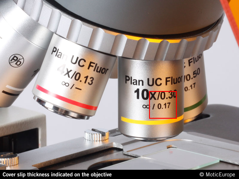 |
| Cover slip thickness indicated on Motic's Plan UC Fluor objectives |
The cover slip has to be placed on top of the sample. A slight pressing of a dissecting needle will help to avoid too much embedding medium (water, etc.) between sample and cover slip. The embedding medium in this case works as an
Thursday, 14 July 2016
Tar spot (Rhytisma acerinum)
The microscopic image shows an infected leaf with sclerotia. A consequence of monoculture.

Just like buildup of mold on pine needles and on the leaves of the willow, an ascomycete (Rhytisma) is the cause of tar spot. From late summer to autumn, round black mold deposits are growing on the leaves of several species of maple, which overwinter on the ground after the falling of the leaves. In spring distribution

Tuesday, 5 July 2016
When you still need a microscope to see Giant structures

Giant chromosomes can be observed in the salivary glands of certain two winged flies (Diptera) and were first observed by Balbiani in 1881 [1]. These chromosomes are oversized and develop from standard chromosomes when specialized cells
Wednesday, 29 June 2016
Rheinberg illumination

An interesting variance of darkfield is Rheinberg
illumination, discovered in 1896 in London by Julius Rheinberg. The major
difference between darkfield and Rheinberg illumination is color. Whereas in
darkfield, the background is black and the subject is white, Rheinberg goes a
step further and creates a colored background and a colored subject.
Monday, 27 June 2016
MOTIC’s New Tablet Solutions: enabling computer-free digital microscopy
Tablets and handheld computer devices are common place in every corner of working life. In schools, clinics or industrial applications, data gets captured, processed and transmitted using Android, Windows or iOS tablet devices.

Bringing this technology and flexibility to proper use in microscopy, Motic’s new Moticam Tablet solutions enable the user to choose the perfect solution for their circumstances. Following on the footsteps of Motic’s recent products to enable computer-free digital microscopy, the Moticam BTU8 and BTU10 feature a fully

Bringing this technology and flexibility to proper use in microscopy, Motic’s new Moticam Tablet solutions enable the user to choose the perfect solution for their circumstances. Following on the footsteps of Motic’s recent products to enable computer-free digital microscopy, the Moticam BTU8 and BTU10 feature a fully
Monday, 20 June 2016
The Dutch live with water
 |
| Micrasterias Rotata and Papillifera | Motic AE31E inverted Microscope | Moticam HD stack |
The Dutch have to live with water, because of their country lying below sea level for the greatest part. What does this have to do with microscopy?
Every year the Dutch are spending billions of euros in dikes, canals, rivers, bridges, locks, waste water purification, water quality monitoring, water research and innovation. Dutch water know how is being applied all over the world.

| Micrasterias Papillifera | Motic BA410E upright Microscope | Moticam 10 |
Thursday, 2 June 2016
Steeling food?
Monday, 30 May 2016
A safe and easy way to operate a Fluorescence microscope for beginners

LEDs are everywhere. This technology is going to roll around also the world of microscopy. For basic illumination purposes, LED technology is already established. Traditional workers will regret the death of the old fashioned Halogen light sources with their emotional high percentage of long wavelengths: think about a candle-light dinner. But LED technology is going to answer this lack of atmosphere by the offer of a large choice of color temperatures. May be this helps.
Wednesday, 18 May 2016
Extremely tiny but very useful ‘hair’

Ciliated epithelium is a type of bodily tissue that is lined with “ciliated” cells, which are basically cells that have small, hair-like protrusions known as “cilia” that can either help the cells move along the tissue or can help debris and waste move along the surface of the cells. Cilia typically move in one direction in a wavelike
Wednesday, 4 May 2016
Color fidelity and fast live image: the new Moticam 1080 Full HD Multi-Output camera is waiting for you!

Turning an ephemeral microscopic image into solid data has become the norm in working with microscopes. But the focus in daily work flow may vary. Sometimes in first instance a speedy live image with true colors for presentation purposes and discussion is needed, and the possible database may be of minor importance. In industrial QC the quantification of saved image data is the key issue. Thanks to its multi-output configuration the Moticam 1080 covers both demands. HDMI and USB signal even can be used to run the camera on HDMI and PC screen in a parallel mode.
Moticam 1080 is dedicated to the presentation and documentation of microscopic results with a clear focus on fast live image and maximum colour fidelity. The impressive 1080 (60P) HDMI live image perfectly fits to the presentation of
Monday, 2 May 2016
Euglenophyta

These organisms are unicellular and live in fresh water. Some of these organisms are photosynthetic, producing their own food, while others are heterotrophic, eating small organisms.
Euglena acus
The Euglena (genus) acus (species) is a type of Protista which lives in fresh water ponds during warm seasons. The Euglena acus produces its food through
Wednesday, 27 April 2016
Motic Images Plus 3.0 – a fresh approach for a powerful microscopy software
The marriage of light microscope and digital camera is a story of success. During the last decade, Motic’ digital products expanded into areas and markets where digital microscopy has been out of reach due to its price perception. One necessary component of this success is a powerful yet easy-to-handle software which covers concordant requirements in medical, educational and industrial applications. From the beginning, Motic Images Plus software supplied useful tools to optimize a fast live image for storage as a still image, image sequence or video. In a second step, a number of quantification tools enable multiple measurements ready to be exported for further use.
Color fidelity is a self-evident expectation on any digital camera. The new Motic Images Plus 3.0 software comes with an interactive White Balance and Background Balance in order to optimize the image background in terms of color temperature or shading. The characteristic of CMOS sensors is responded by a Color Correction tool.

A calibrated Scalebar in the live image helps for a first estimate of size and can be saved as an overlay; a variable Grid may replace the pattern of various counting chambers. But also Interactive Measurements are now possible within the Live image. The necessary calibration slide is integral part of Motic’s All-in-One camera concept and causes no additional costs.

Motic Images Plus 3.0ML will start shipping as standard with the new upgraded Moticam Plus cameras (that will come out in the next coming months) as well as the new multi-function HDMI camera, the Moticam 1080.

As is normal practice with Motic, each end-user can upgrade their existing Motic Images Plus 2.0 to the new 3.0 anytime, anywhere, and free of charge. This sleek, new and thought-out software package is running with an identical interface on Windows, Apple or Linux operating systems and certainly empowers your microscope to be a more efficient scientific tool. Even more as Motic Images Plus 3.0 is a real Multilingual software; a change of language is done within a second.
Click here to download the new Motic Images Plus 3.0.
Monday, 25 April 2016
This is not a pink cube

There is controversy on whether cobaltocalcite
is a mineral on its own or if it is not. It can be described either as calcite
mineral containing cobalt or as spherocobaltite containing calcium. It surely is a calcium and cobalt carbonate and, as it happens with spherocobaltite, it is
pink, vitreous and it always grows incrusted on a matrix.
Wednesday, 13 April 2016
The retina, a bio-chip on the back of the eye

The retina which is located on the back of the eye, is composed of very closely spaced light-sensitive cells that are in connection with the brains via the optic nerve. The light signals that reach the light-sensitive cells, are transferred to the brains via the optic nerve. The brains takes these signals and translates them into the picture what is happening before our eyes.
Friday, 8 April 2016
Quick setup guide for upright microscopes
SWITCH ON THE MICROSCOPE
Before switching on the microscope, plug-in the microscope to the power supply (1). Once this is done, switch it on (2) and gradually increase the intensity control (3).

INTERPUPILLARY DISTANCE ADJUSTMENT
Before adjusting the interpupillary distance, place the sample on the specimen holder, and select the 10X objective (4).

Tuesday, 5 April 2016
Some ideas about the FIELD of VIEW
To judge the performance of a microscope, the FIELD of VIEW (FOV) is a criterion of interest. What is this “FIELD of VIEW”? In a technical sense, we are talking about the diameter of the INTERMEDIATE IMAGE, created by the objective or, in infinity systems, by the cooperation of objective and tube lens. This intermediate image is enlarged by the eyepiece, acting as a magnifying glass. The labelling of the eyepiece shows the actual FOV in combination with its magnification, e.g. 10X/23 for a standard 10X eyepiece with FOV 23:


Especially in stereo microscopy, where large samples come as standard, a large FOV is appreciated for a better sample overview before going into details. But how much “more” do I see at a glance in a FOV 23, in comparison with a FOV 20? Is it worth to choose a more expensive model, or is the less expensive solution sufficient?


Finite and infinite optical system. In the Infinity Concept please note the parallel rays between objective and tube lens.
Especially in stereo microscopy, where large samples come as standard, a large FOV is appreciated for a better sample overview before going into details. But how much “more” do I see at a glance in a FOV 23, in comparison with a FOV 20? Is it worth to choose a more expensive model, or is the less expensive solution sufficient?
Wednesday, 30 March 2016
A cactus takes breath at night

Most plants have their pores (stomata’s) open during the day to take in carbon dioxide, and use sunlight as a catalyst for the photosynthesis. But in the desert, plants with pores open during the hot days, lose much water through evapotranspiration. So, succulents use a modified version of photosynthesis called CAM (Crassulacean Acid Metabolism).
CAM plants open their stomata’s only at night when it is cooler so there is less evapotranspiration. Because there is no sunlight to act as a catalyst, carbon dioxide is stored as an organic acid, principally Malic Acid (C4H6O5). Carbon dioxide is
Thursday, 24 March 2016
Azurite to malachite

Tuesday, 22 March 2016
You can find it on rotting fruit

Rhizopus is a genus of common saprobic fungi on plants and specialized parasites on animals. They are found on a wide variety of organic substrates, including "mature fruits and vegetables", faeces, jellies, syrups, leather, bread, peanuts and
Wednesday, 9 March 2016
Magnification on screen
One of the most frequently asked questions about digital imaging is:
what is the total magnification showed on the screen?

To answer this question, we need to know four things:
Tuesday, 1 March 2016
Xanthidium armatum ‘inverted’

The use of an inverted microscope for observation of phytoplankton, actually belongs to the standard. The image which is shown here, has been taken with the Motic Inverted Microscope AE31E. A 35 mm cell culture petri dish with a high precision glass bottom was used. This combination has some advantages over the use of an upright microscope:
Friday, 19 February 2016
MITOSIS: Multiplication by Division
Our body contains trillions of cells (thousands of millions) but everything started from a single cell. This unit cell has the amazing ability of dividing into two, and then four, and then eight and so on, in time becoming a complete organism. The process through which cells divide and divide as to multiply in number and become a complete organism, is known as Mitosis.

During Mitosis, the parent cell divides to form two genetically identical daughter cells. In order to assure a perfect copy of the genetic material in the daughter cells, the parent cell has to duplicate the chromosomes first; this step is called the

Wednesday, 17 February 2016
Feulgen stain, still indispensable for microscopic research

Wednesday, 3 February 2016
Painting with crystals
Preparing a normal or time lapse video of a crystallization process under the microscope is not easy. Focusing problems, formation of too big crystals and getting the interesting part right under the objective, often gives you a headache.
With the technique described below, most of these headaches can be overcome:
With the technique described below, most of these headaches can be overcome:
Thursday, 21 January 2016
Finding the pot of gold at the end of the rainbow

Wednesday, 20 January 2016
Ants, the world’s barely visible largest biomass

Ants are a group of colony-forming social insects, which belong to the order of Hymenoptera. Ants have been able to adapt to very different habitats; where they occur, ants are the dominant life form on the bottom. It is estimated that the total biomass of ants is greater than that of any other animal species on earth.
Thursday, 7 January 2016
Just stones?

Subscribe to:
Posts (Atom)