Wednesday 20 August 2014
A family existing over 500 million years
Foraminifera are amoeba-like, single-celled protists (very simple micro-organisms). They have been called 'armored amoebae' because they secrete a tiny shell (test) usually between about a half and one millimeter long. They get their name from the foramen, an opening or tube that interconnects all the chambers of the test. Fossilized tests are found in sediments as old as the earliest Cambrian (about 545 million years ago) and foraminifera can still be found in abundance today, living in marine and brackish waters. Although each foram is just a single cell, they build complex shells around themselves from minerals in the seawater. These shells have accumulated in layers of sediment below the seafloor of the open ocean and in regions where the ocean once flooded the continents for millions of years. By examining the shell chemistry of these ancient forams, scientists can learn about Earth's climate long before humans ever walked the planet and get insight into how climate changed in the past.
Sources: Smithsonian National Museum of Natural History, NERC Science of the Environment
Monday 11 August 2014
Experiment: Observation of onion epidermial cells
Material
- Raw onion
- Slides and cover glass
- Scalpel and forceps
- Water and dropper
- Filter paper
- Methylene blue stain solution
- Microscope, Digital microscopy camera
Procedure
Place a couple of drops of water in the center of the slide.
Using the scalpel, slice the raw onion. Cut one of the onion
rings into 6mm sections.
By using the forceps, gently remove the skin from the inner side of the onion.
Place immediately the skin on the drop of water to avoid it curls up. Gently place the cover glass. If necessary remove the water excess with filter paper.
Prepare a new sample by placing a couple of drops of diluted methylene blue solution in the center of a slide. Place a new onion skin on this solution and cover with the cover glass, as before. If necessary remove the staining solution excess with filter paper.
Observe the slide again under the microscope using the same low-power and high-power magnifications.
Observe the pictures and identify the shape and pattern of the onion cells.
Compare the pictures and find the differences on the sample before and after staining.
The onion epidermis, because of its simple structure and
transparency, it is often used to introduce students to plant and cell anatomy.
Students should be able to distingue the rectangular shape
of the individual cells, which are perfectly lying side by side, forming the
typical epidermis pattern.
After the staining students should be able to identify
cellular structures (mainly the cell walls and nuclei) which are almost
indistinguishable on the live samples.
Staining enhances the optical contrast of these structures making them
visible.
Wednesday 6 August 2014
Algae have sex too!
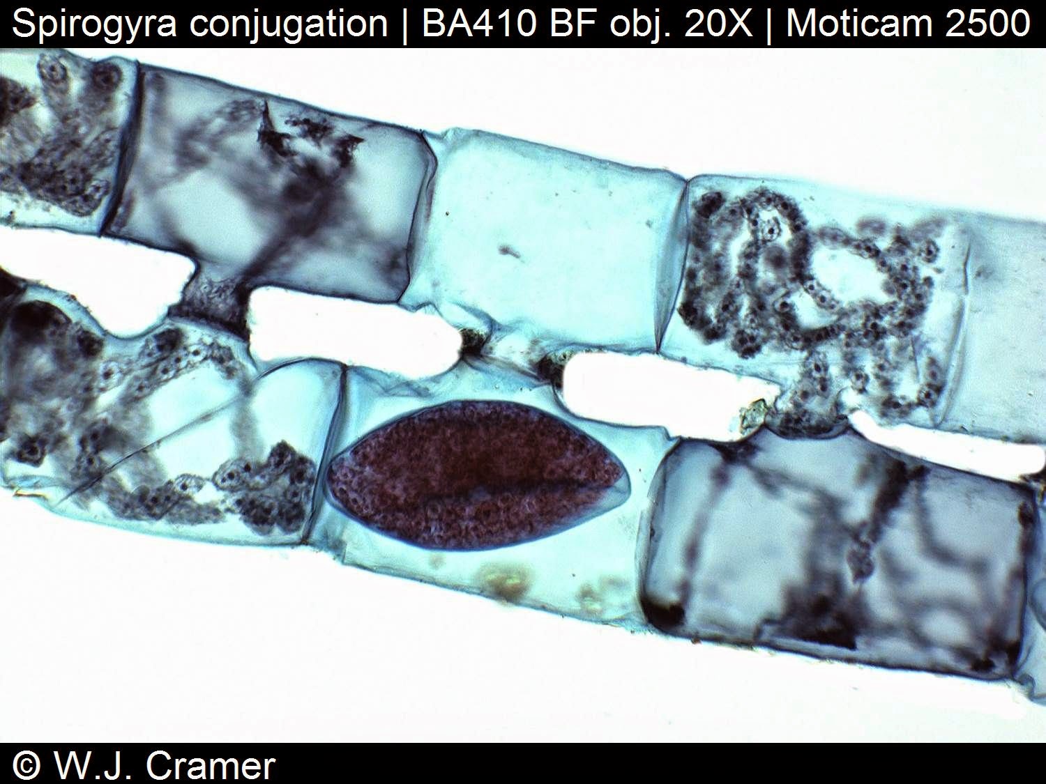
Algae have sex too! (at least some of them, though who knows if they enjoy it?) Amongst the Zygnemaceae, the process of conjugation consists of the joining together of the contents of two haploid cells from (usually) different filaments.
Conjugation in Spirogyra: the filaments show the typical results of scalariform ('ladderlike') conjugation. Two filaments lie parallel, and outgrowths from each filament grow towards each other, and then fuse to form a conjugation tube. The conjugation tubes allow the contents of one cell to transfer to the other. The dark oval shapes are diploid zygospores, formed after nuclear fusion between the two haploid cells. They form in only one of the two filaments. Sometimes more than two filaments take part in conjugation like a 'menage a trois'.
Spirogyra is a filamentous green alga in which the chloroplast has a characteristic spiral shape. In one of the photographs, you can see the chloroplast coiling against the outer edge of the cells. The numerous small round blobs along the edges of the chloroplast are the pyrenoids. The larger, faint blobs (they look like out-of-focus regions) that take up most of the volume of the cells in the filaments on the right are the nuclei, which are suspended in the interior of the cells.
Pyrenoids occur in many of the algae and are associated with the chloroplasts. Some of them are known to contain Rubisco, the enzyme that catalyzes the incorporation of inorganic CO2 into carbohydrate (Graham and Wilcox, 2000). In these algae, pyrenoids probably function to fix carbon. In other algae, pyrenoids are the sites of carbohydrate (typically starch) storage. Starch and iodine react to produce a deep blue- black color, so staining a thin algal prep with iodine will indicate the presence of pyrenoids.
Sources: Algalweb, MadSci Network
Subscribe to:
Posts (Atom)


