Monday 25 April 2022

Mercury, the only liquid mineral

The classic definition of “mineral” is, in general terms, a mineral is an element or a chemical compound that is normally crystalline and that has been formed because of geological processes. (Nickel, 1995).

But with the progress of mineralogical and chemical science, this "general" definition has been refined. Thus, Donald Peck proposes to define what a mineral is as an element or chemical compound that:

1. has a more-or-less constant composition -with allowance for substitutions in specific crystallographic sites in continuous series.

2. is usually a solid with an ordered three-dimensional array of ions and molecules in its crystal structure. -A few exceptions are approved for well-characterized amorphous substances.

3. is formed by natural geologic processes and without human or other biologic intervention. Exceptions are made for organic materials that have been changed and crystallized by geologic processes.

4. is not a mixture of two or more blended substances.

And, he adds that the most important thing is that it has been exhaustively studied and characterized by mineralogists, declared unique in its composition and structure, and that the original specimens (holotype) studied are deposited for conservation in a "serious" museum (Peck).

If we take these requirements into account, what is mercury? an ore?

At room temperature, it is a silvery, shiny liquid with a high surface tension that makes it easy to form balls and when they fall on a surface it is difficult to collect them... Who has not broken a mercury thermometer (actually prohibited)?

As it turns out, mercury is officially classified as a mineral species. It has a constant composition, it is not a mixture, but it is not solid, nor does it have an ordered three-dimensional array: Mercury is considered an element only for historical reasons and because it is distinctive in its chemical and physical properties. Because of its age.

The name of this liquid metal comes from the Roman god of merchants and messenger of the gods. Its current chemical symbol is Hg, from the word hydrargyrum, a Latinized form of the Greek term ὑδράργυρος (hydrargyros), which is a compound word meaning "water-silver", which seems obvious. The symbol ☿, which represents the planet Mercury, was used by alchemists to represent this liquid metal, an ingredient to "synthesize" gold.

Mercury does crystallize (solidify), in the form of rhombohedral crystals, when we cool it to −38.83 °C. In the Almadén mines (Ciudad Real, Spain), which at the time was the world's largest producer of mercury, liquid mercury can appear as pockets, in fissures (it already meets another requirement: formed by natural geological processes) and associated with cinnabar, mercury sulphide (HgS). The main ore of Hg is this intensely red mineral, also used in the past as a pigment and known as vermilion.

Fig 1 - Cinnabar crystal with quartz, FOV 5 mm. Motic PlanApo 5X with stacking. Collection & photo: J. Rosell. 

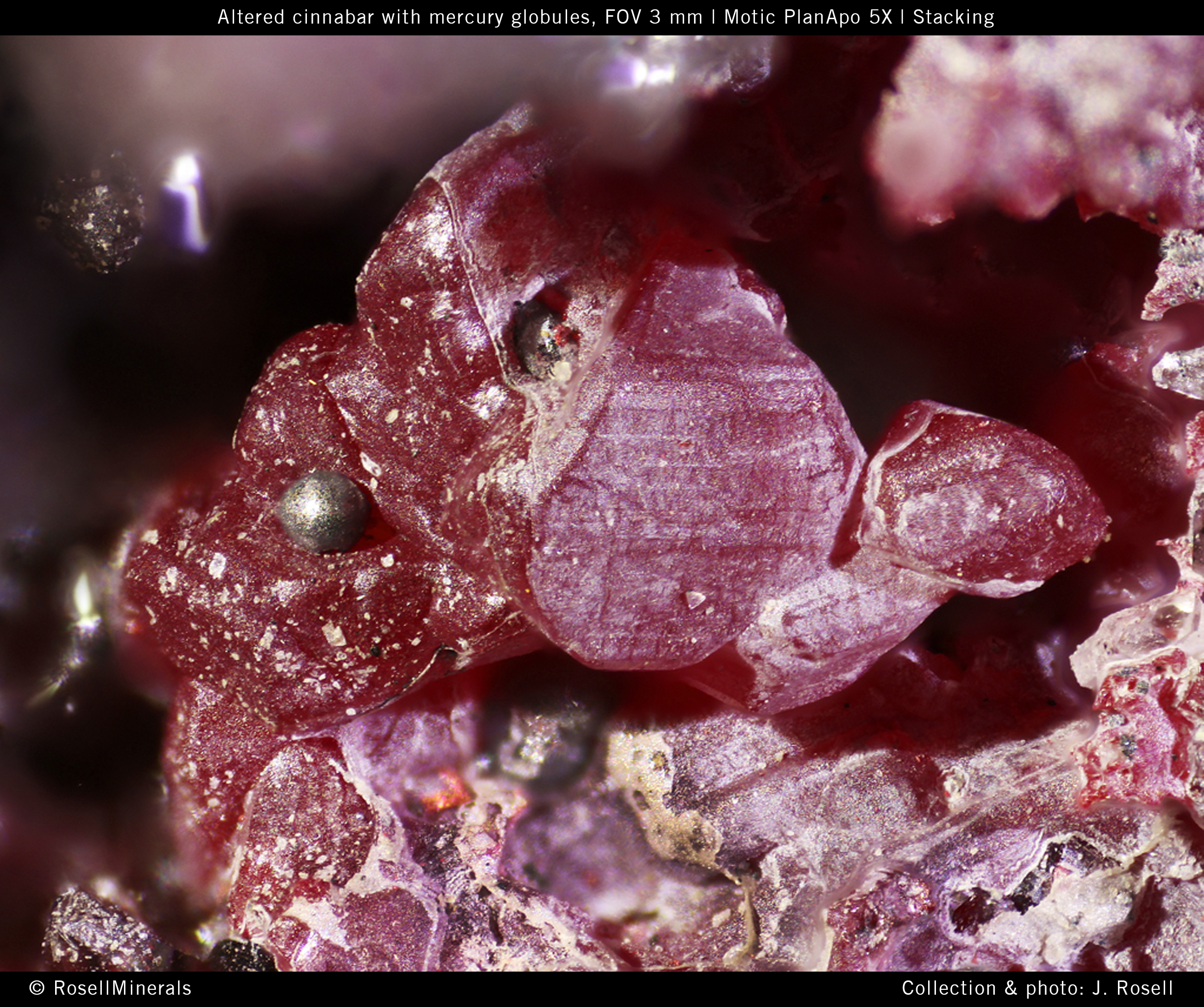
In the photographs, we can see small balls of mercury that remain attached to the rock with red cinnabar, dolomite, and quartz. These globules appear when the rock is broken and remain attached to it unless we give the piece strong blows or pass a brush. Interestingly, if we try to deposit mercury on the rock from a bottle of liquid mercury, we will not succeed, and the mercury will slip on the piece.

Fig 2 - Brilliant globules of mercury on cinnabar (red) and quartz, FOV 4 mm. Motic PlanApo 5X with stacking. Collection & photo: J. Rosell. 


Whenever we handle minerals, we must be careful to wash our hands, and even more so if it is cinnabar. Mercury is toxic. It is said that having it in your hand is dangerous, let's say that the vapours emitted are scarce (Hg vapour pressure at 20ºC is 0.16 Pa, water is 2300 Pa). What is dangerous is when Hg reaches food chains, where it penetrates as organic compounds generated by bacteria (mainly methylmercury), although curiously, it may be microorganisms that clean the seabed contaminated with mercury.

Fig 2 - Altered cinnabar with mercury globules, FOV 3 mm. Motic PlanApo 5X with stacking. Collection & photo: J. Rosell. 


References
Nickel, Ernest H. (1995): “The definition of a mineral”. Canadian Mineralogist, vol. 33, pp. 689-690.
Peck, Donald: What is a mineral? Mindat.org  [https://www.mindat.org/a/what_is_a_mineral]


Friday 22 April 2022

Spring bouquets

In a small fen, located in a heather area somewhere in the south of The Netherlands, during a sunny period in spring, we saw a bloom of Dinobryon divergens. Dinobryon is very common and is one of several types of golden algae. The presence of golden algae can be an indication of reasonable water quality.

Under the microscope, these algae provide a nice colorful picture. They have the shape of a bouquet of flowers. You would almost say 'they are spring bouquets'. They can move freely with their cilia, which are clearly visible at the larger magnifications in the video. The video was made using the Motic AE31E inverted microscope and the Moticam S6, a member of the new S Series of Motic.


Saturday 16 April 2022

The art of preparing botany samples

Fabrication of a botanical preparation is not that easy.

It is a lengthy and laborious activity to make a botanical preparation as is shown in the images. It is quite an art to maintain the natural shape of the material, to cut the section as thin as possible, color it, and keep the loose parts of the flower bud in place. We will not go into too much detail about the necessary procedures to achieve a good result. All this has extensively been described in the relevant literature.

Briefly summarized, the following steps are generally taken:

Fixation of the plant material with for example alcohol, an alcohol glacial acetic acid mixture, or a formaldehyde solution. This must be done in such a way that the material does not lose its natural shape due to shrinking. After fixation, the fixative must be washed out.

If necessary, the fixed material can be stored longer in alcohol, glycerin, and water mixture.

In the botanical microtechnique, the embedding of the fixed material in paraffin is most commonly used before cutting very thin slices out of it. Before embedding, the water must be removed from the material with a solvent such as alcohol.

Before embedding in paraffin, the material can be clarified if necessary using certain solvents.

The material now enclosed in cubes of paraffin, is cut into micrometer thin slices, the so called coupes, using a slide or a rotary microtome. These are equipped with razor-sharp knives.

The resulting coupes are attached to an object glass using a special adhesive. The coupe is stretched by careful heating so that it lies as flat as possible on the object's glass. The paraffin is then washed out with a solvent.

Then the various structures in the coupe are colored. There are many coloring methods available, which make it possible to distinguish structures in the coupe with their typical functions. After coloring, the coup must be made free of water before the next step.

In order to preserve the preparation for ‘eternity’, the coup is embedded in a mounting agent. The mounting agent is usually a natural or synthetic resin.

During embedding, the coup is covered with a coverslip, heat may also be added to allow the mounting agent to flow properly. The trick is to avoid the inclusion of air bubbles.


Fig. 1 - Male pine cone seen under the Motic Panthera C microscope, Plan UC 2X, Moticam ProS5 Plus

Fig. 2 - Male pine cone seen under the Motic Panthera C microscope, Plan UC 20X, Moticam ProS5 Plus

Fig. 3 - Male pine cone seen under the Motic Panthera C microscope, Plan UC 40X, Moticam ProS5 Plus


Fig. 4 - Male pine cone seen under the Motic Panthera C microscope, Plan UC 60X, Moticam ProS5 Plus

© www.willemsmicroscope.com

Thursday 14 April 2022

Centropyxis: an unknown in the biofilm, an opportunity to discover.

The “Escalón” cave, located in the Asón Valley in Cantabria (Spain), offers great possibilities for carrying out astrobiology work, and the company Astroland Agency has installed an operations base there, the “Ares Station”. From it, the sampling and recognition of biofilms have been organized, these grow on the humid walls of the cavity, in which these facilities are housed.

From the "Ares Station" different protocols have been tested to carry out research related to the study of the biofilms that cover the limestone rocks around the cave, which in this case, and unlike what happens in other cavities, are constituted, fundamentally, by cyanobacterial communities with species that are currently being investigated, and that are of great interest, because exceptionally scarce organisms have been found in them, and in some cases, not yet described.

Fig.1. The appearance of the cyanobacterial masses where this amoeba, Centropyxis sp. (aerophyla group) in the surroundings of the Ares station. Gloeobacter violaceus and Schizothrix sp. are some of the cyanobacteria among which it seems to be found most frequently.

These biofilms in the environment of the Ares Station have been revealed as a treasure of microbial biodiversity, and although most are made up of a complex and varied tapestry of cyanobacteria, they are also home to other microorganisms of great interest. Among them, some testate amoebas such as Difflugia alhadiqa, were discovered and described for the first time by our colleagues Carmen Soler-Zamora, Miguel González-Miguéns, and Enrique Lara, very recently, in the Hundidero cave, Montejaque, (Málaga, Spain). Another microorganism - that we show here and will have to be investigated more deeply - corresponds to a complex of amoebas of the genus Centropyxis, very close to the group of Centropyxis cassis, which present an enormous taxonomic difficulty.

Fig. 2. Naiara and Irene are high school students and they participate in this research work by assembling and preliminary observing a biofilm sample using equipment provided by Motic, the SMZ-168-TLED stereomicroscope.

The genus Centropyxis described by Stein in 1857 includes testate amoebas with a more or less discoidal and slightly flattened shell, with a flat or concave ventral side and a convex dorsal side. One of the most remarkable characteristics of this genre is that the opening of the shell, of very varied contours, is displaced towards one end.
A group of these fundamentally aquatic amoebas may have spines in a very varied number on their periphery, but this is not the case at hand. The species we are referring to here does not present them, although, like any of the representatives of the genus, its cover is covered with mineral particles, cemented by an organic matrix.

Fig.3. Centropyxis sp.(aerophyla group) among the cyanobacterial biofilm with Aphanothece saxicola dominance. Photographs taken at 400x magnification with the epifluorescence technique, with the FLED module and the Motic 40X/0.65/S (WD 0.6mm) dry objective. Equipment used: Motic Panthera CC trinocular.

More than 135 species and many varieties have been described, but many descriptions are currently being revised. Morphological recognition is insufficient to be able to accurately determine species with precision, which is only possible using genetic sequencing techniques.

There is currently an interesting debate about the identification of the species of the Centropyxis cassis complex, Centropyxis aerophila and Centropyxis constricta, which it is not appropriate to comment on in this publication, but it does seem clear that despite the fact that the morphology of our species is closer to Centropyxis cassis due to the shape of the theca, its dimensions are far from being those of the latter, which in C. cassis oscillate between 54×60 µm wide and 64×75 µm long. In the few specimens found among the biofilm of the cave, the dimensions range between 35–40 µm wide and 50–58 µm long and 20–15 µm in diameter, in the major and minor axes, respectively.

The form of C. constricta is very reminiscent of that of the species discussed in this article, but its size is much larger (about 100 µm) and its habitat is completely different - it lives in bright and cool areas with Sphagnum.

Fig.4. Centropyxis sp.(aerophyla group) in another view among the cyanobacterial biofilm with a domain of Aphanothece saxicola. Photographs taken at 400x magnification with the epifluorescence technique, with the FLED module and the Motic 40X/0.65/S (WD 0.6mm) dry objective. Equipment used: Motic Panthera CC trinocular.

From C.aerophyla, with almost spherical contours and larger dimensions, it seems to be further away both because of its shape and size and because of its habitat.
Everything seems to indicate that the species we show here is different and that it could be a new species. All this will have to be confirmed in subsequent sampling and research, in which the sequencing of its genetic material will be key.

Fig.5. Observing the samples of Centropyxis sp. (aerophyla group) with the FLED module and the Motic 40X / 0.65 / S (WD 0.6mm) dry objective. Students of 1º of Baccalaureate of the subject Scientific Culture in the “IES Escultor Daniel" (Logroño, Spain).

Possibly Enrique Lara's team from the “Real Jardín Botánico” of Madrid and other experts such as Edward Mitchell or Foissner and Korganova, who have worked on this genre, can make their contributions and comments.

In any case, it is a relevant finding and further proof of the interest presented by these barely studied biofilms.

All the photographs have been taken at a magnification of 400, with bright field and epifluorescence techniques, with a Motic Panthera CC trinocular equipment and come from the samples collected inside the Escalón cave, in the Ares Station environment. It is a very dimly lit area where the Astroland Agency is developing an approach to learning about Mars in its astrobiological project.

Fig.6. Centropyxis sp.(aerophyla group) from a fresh sample of the cyanobacterial biofilm. Photographs taken at 400x magnification with the epifluorescence technique, with the FLED module and the Motic 40X/0.65/S (WD 0.6mm) dry objective. The opening of the theca and the contour of this amoeba can be seen very well, as well as some cells of the Gloebacter colonies in yellow. Equipment used: Motic Panthera CC trinocular.

Today this amoeba sees the light from the darkness of the cave where it was found for those who come to read this publication and for the students of the sculptor Daniel High high school in Logroño. They have participated in these observations handling the Motic equipment - both microscopes and stereomicroscopes - learning how to handle this equipment both with conventional brightfield lighting and with the FLED epifluorescence module, essential for identifying and seeing the mineral particles that make up the theca of this amoeba.

Wednesday 13 April 2022

Filming tiny animals in a drop of water

With a stereo microscope such as the Motic SMZ-171 and a camera such as the Moticam 1080, you can make excellent photos and videos of tiny aquatic animals swimming around in a small amount of water (sometimes in a single drop only) which is in a Petri dish or on an object-glass. The illumination of the object is essential here.

A Motic darkfield attachment and side lighting with two flexible optic fiber LED lamps were used for this video. The trick is to suppress the glare caused by the reflection of light from the water drop as much as possible. This was not entirely successful here, because indirect side-illumination was not used, whereby the light rays are first reflected on a white surface before hitting the object. An opal filter could also have been used.


The video starts with the title ‘Daphnia’. We are dealing here with the Simocephalus vetulus. This water flea has a relatively small head, clearly demarcated from the shield by a cavity on the dorsal side. The maximum height of the shield is behind the center. The green algae that we see are called Spirogyra, so named after the spiral arrangement of the chloroplasts.

Wednesday 6 April 2022

Art, design and crystals

In her studio in the Port of Rotterdam, the innovative artist and designer Liesbeth Bulk is experimenting with the growing of crystals on every day and design objects. She tries to combine the beauty of crystals with the structure of objects. Mastering this artistic and technical challenge is still at an initial stage. Until now, trials have been carried out with sugar crystals. When we look at the picture taken with a Motic stereo microscope, it is not surprising that artists are fascinated by the beauty of crystals and start experimenting with them.  

Fig. 1 - A sugar crystal under the SMZ-71 Motic Microscope 

Liesbeth is a designer with a fascination for nature, she also creates bespoke glass panels with enclosed natural elements. With profound knowledge of plants, the flowers, leaves, and twigs are collected in the wild. Searching for a way to merge the lush plants with monumental, architectural characteristics, she started experimenting with the forgotten technique: pressed flowers.

Fig. 2 - A small part of Liesbeth’s art lab. 

Liesbeth Bulk (1968) studied garden design at the Horticultural College in Boskoop, The Netherlands. In 1998 she completed her studies at the Sandberg Institute (post-academic course at the Gerrit Rietveld Academy Amsterdam) with a Master in fine arts. For examples of her work use the links below.

Friday 1 April 2022

Recovered from the bottom of the sea

These three heavily corroded aircraft engines were fished up by Texel fishing boats on the North Sea. All three of them are from planes that crashed at sea in World War II. The one in the middle is a 14 cylinder radial engine labeled Pratt & Whitney, manufactured in the USA. The Liberator and the Halifax were equipped with such an engine in the war. The other two engines are labeled Bristol Hercules and were manufactured in Britain. The heavy and medium heavy bombers of the RAF were equipped with this type of engine. It is different from other engines. It does not have any intake and outlet valves but a working cylinder with ports in the 2-stroke principle. 


Fig. 1 - Museum Kaap Skil, Oude Schild, Texel
Texel is n island in the North Sea, in the north of The Netherlands.

The microscopic images show serious corrosion to the aluminum alloy of a certain part of the engine, caused by the aggressive seawater. The corrosion product shows fluorescence when exposed to UV light. These are compounds that are formed by a chemical reaction between the metals present in the alloy and the seawater.

Fig. 2 - Corroded aluminum alloy aircraft engine part under an SMZ-171 microscope and a Moticam 1080 BMH stack  

Fig. 3 - Corroded aluminum alloy aircraft engine part under a BA410E microscope PlanFluar 10 X (DAPI filter) and a Moticam 10 stack.  
© www.willemsmicroscope.com