

Tuesday, 30 December 2014
Optical fibers in sponges?
Friday, 19 December 2014
Too many mouths?

Stomata, are the mouths of plants. Plants breathe through them, just as we do with our lungs. They are not visible to the naked eye, and a plant therefore needs quite a lot of them. However, there must be
Wednesday, 3 December 2014
Hair? Where?
True hair is found only in the Class Mammalia, and there is really no such thing as an absolutely hairless mammal. Even whales (at least some of them) have rudimentary hairs here and there. Some other animals have hair-like structures, but if you have real hair, you're a mammal.

Hair serves many functions. The most obvious is to serve as insulation, but it's also used to provide camouflage (for example, the spot pattern
Hair serves many functions. The most obvious is to serve as insulation, but it's also used to provide camouflage (for example, the spot pattern
Wednesday, 19 November 2014
Workhorse Aspergillus Niger
Aspergillus Niger is a fungus and one of the most common species of the genus Aspergillus. It causes a disease called black mold on certain fruits and vegetables such as grapes, onions, and peanuts, and is a common contaminant of food. It is ubiquitous in soil and is commonly reported from indoor environments, where its black colonies can be confused with those of Stachybotrys (species of which have also been called "black mold").

Aspergillus Niger represents the most efficient, highest yielding bioprocess for the production of citric acid in practice. This process is a model for other filamentous fungal fermentation processes
Monday, 17 November 2014
Autumn - Time for Mushrooms
The autumn arrived and with it, the while mushrooms. Do you want to see how they look like under a microscope?
We used a SMZ-171 TLED and a Moticam 5 to observe some particularities on Lactarius deliciosus, commonly known as the Saffron milk cap, Red pine mushroom.

These mushrooms grows under the
These mushrooms grows under the
Wednesday, 12 November 2014
Ladybug ‘salto mortale’
Friday, 31 October 2014
How to avoid repairs
This time we would like suggest some easy steps that will help you maintain the microscope in perfect conditions, and avoiding possible repairs. You might already know them, but we think it is good to refresh your memory from time to time.
Before using the microscope
Read the instructions manual of the instrument carefully.
Never attempt to disassemble the parts of the instrument, other than the ones described on the manual.
Dust and dirt are two of the main causes for malfunction in a microscope.
Before using the microscope
Read the instructions manual of the instrument carefully.
Never attempt to disassemble the parts of the instrument, other than the ones described on the manual.
Dust and dirt are two of the main causes for malfunction in a microscope.
Thursday, 23 October 2014
And the winner is...the new BA410 Elite with Apochromatic objectives!

The apochromatic correction of a lens system sine dubio defines the ultimate level of optical performance. Ernst Abbe (1840-1905), one of the fathers of modern optics, created this word derived from the Greek origin, meaning “free of colours”. Of course Abbe was talking about the colour abberations of simple optical systems. These abberations are caused by the dependency of refractions on
Wednesday, 22 October 2014
Smoking
Lung cancer is the most common form of cancer caused by smoking. More than 80% of cases of lung cancer are due to smoking.
Cigarette smoke contains many chemicals that interfere with the body's method of filtering air and cleaning out the lungs. The smoke irritates the lungs and leads to overproduction of mucus. It also paralyses the cilia - tiny hair-like structures that line the airways and clean out dust and dirt. Paralysis of the cilia means mucus and toxic substances accumulate, resulting in congestion of the lungs.
This extra mucus means smokers are more likely to suffer from chronic bronchitis and what is known as 'smoker's cough'.
Cigarette smoke is one of the best known triggers of asthma. When people suffer from asthma their inflamed air passages, which are very sensitive, narrow when exposed to cigarette smoke. This causes an asthma attack.
Long term exposure of the lungs to the irritants in tobacco smoke destroys the normal lung structure. The elastic walls of the small airways within the lungs are broken down. This reduces the amount of lung tissue available for the transfer of oxygen from the air to the blood. This condition is called emphysema. Some degree of emphysema is found in almost all people who are long-term smokers, however the severity will vary depending on the amount of cigarettes smoked, and the number of years the individual smokes.
Damage to the lung tissue is irreversible. Emphysema can be prevented by not smoking, avoiding anything that will irritate the lungs such as dust and cold air, and ensuring any chest infections such as flu and bronchitis are treated properly.
Source: Quit org. au.
Source: Quit org. au.
Friday, 10 October 2014
An itching subject

Fleas extract and consume the blood of host animals in order to survive. Neither cat nor dog fleas leave the host voluntarily and will typically remain with one host throughout their lifespan. However, if dog fleas are forcibly removed from their host, they will locate a new host or return to the original host if possible.
The life cycle of the flea is composed of the egg, larval, pupal and adult stages. Cycle length ranges from several weeks to several months and is largely dependent upon environmental conditions. Fleas lay between four to eight eggs after a meal, with the highest concentrations of laying occurring within the last few days of the female’s life. Unlike the eggs of some other parasites, flea eggs are not sticky and usually fall to the ground immediately upon being laid. Flea eggs hatch into larvae within one to 12 days. Flea larvae are approximately 3 to 5.2 mm long and are semitransparent white in color. The larval stage lasts from four to 18 days, after which larvae spin silken cocoons and enter the pupal stage. The pupal stage may be complete within three days, or it can last as long as one year.
Adult fleas begin searching for food when they emerge from the pupal stage. While fleas are noted for their jumping abilities, they will remain stationery when a suitable host is located. Females begin laying eggs within 48 hours of the first feed, thus beginning the life cycle again.
Source: Orkin
Thursday, 9 October 2014
Refined and optimized…Motic’s new inverted microscope, the AE31 Elite, is ready!

The idea of an inverted microscope bears further advantages. The beginner’s step into the world of small things is as easy as with a stereo microscope: No need for sample preparation, no sectioning, no need for staining: just take a sample from your rain gutter. It is easy to find algae, paramecia, rotifers, even water bears with their astonishing constancy of body cells. For beginners, the intrinsic true sided upright image (which finally is dedicated to professional micromanipulation work) helps in orientation.
The AE31 Elite is a professional inverted microscope with an extended focus on practical aspects. The big brother of Motic’s entry level model AE2000 also implements the AUTO ON-OFF via IR sensor for energy-saving and safety reasons. The LIGHT MEMORY function, based on the encoded 5-fold nosepiece, helps to keep the proper illumination intensity: Once set up for each objective, there is no need to readjust the illumination when changing the magnification.
To harmonize the optical setup of the CCIS© Infinity concept, the AE31 Elite works with the newest generation of LWD Plan Achromatic lenses: Only one Phase ring is needed for Phase 10X up to 40X; the objective Phase 4X for fast screening is available as an option. These features establish the AE31 Elite as a microscope platform for live cell biology.
The tube lens of the AE31 Elite has been harmonized with the Motic’s BA series of upright microscopes. So all accessories like c-mounts, additional eyepieces, etc. can be used without restriction. The fully corrected intermediate image is ready for digital access. Fluorescence as an upgrade option remains a strong feature and separates the AE31E from the smaller model AE2000.
The AE31 Elite continues Motic’s approach for a balanced selection of professional yet affordable instruments for most aspects of microscopic work in biomedical labs.

Tuesday, 7 October 2014
What is Phase Contrast and when to use it?
Most of the Biological samples are quite transparent, characteristic that turns difficult its visualization under a Brightfield microscope. To overcome this, scientists frequently treat the samples with different staining solutions in order to enhance their contrast.
Nevertheless, this option cannot be considered when working with living cells, since the chemical toxicity of the staining solutions would kill the cells. This is a particular issue for Microbiologists, who frequently observe living cells (usually in culture), microorganisms, and thin tissue slices.
The Phase Contrast technique, is an easy and smart way to enhance contrast on transparent specimens, and therefore allows the examination of living cells in their natural state.
A phase contrast microscope enhances the change in phase of the light going through the sample and thereby causes a difference in brightness. To see which components you will need to use this technique, and how to set up them on your Motic microscope, please see our Simple Phase Contrast video.
The images below show thin slices of a Rabbit testicle, where the spermatogenesis process can be followed. Both pictures(100X magnified) were taken with a Moticam 5 mounted on the Motic AE2000 inverted microscope. On the left, the sample visualized in Brightfield and on the right with the Phase Contrast technique.

Friday, 26 September 2014
The cover slip - With or Without You
The uncommon yet simple rule “The sample defines the microscope” certainly helps to choose the appropriate microscope hardware for a defined application. Biomedical tissue sections or smears require a combination of upright microscope with transmitted light, while cell cultures (inverted plus transmitted light), flat and polished metal specimen (upright or inverted plus incident light), birefringent materials (upright POL plus transmitted light) or 3-dimensional biological beings (stereo microscope) request different microscope setups.
But there is a wider understanding of this guideline necessary to get the best possible image quality. A section of an organ tissue, a smear of a human mucous membrane, adherent or floating cells in microbiology: They all need a carrier or vessel to be evaluated under the microscope.
Sections or smears are fixed on a glass slide, in most cases conserved by a suitable embedding medium and fixed by a cover slip of defined thickness. Native biological samples (the leaf of a moss, pollen grains) can be placed in a drop of water with a cover glass on top. The proper microscope hardware in these cases is an upright model, while the sample additionally asks for a careful preparation.
Standard transmitted light objectives are designed for a 0.17mm cover slip between naked sample and front lens. This indication can be read on the objective sleeve. Cover slips are industrial products with astonishing manufacturing tolerances (a stupendous fact in the world of optics where precise distances are essential!). The standard cover slips have a thickness of 0.13-0.16mm, taking the add-on layer of embedding medium or water into account to follow the 0.17mm rule.


The higher the Numerical Aperture of an objective, the higher the sensibility for deviations from this value is. A 4X/0.10 or 10X/0.25 Plan Achromat may be used with or without cover glass without obvious visual effects. But taking a 40X/0.95 from Motic’s new PLAN APO series, we seriously have to take care for the best possible preparation.

Special cover slips with a tolerance of 0.17+/- 0.005mm are the best solution for that kind of demanding optics. No need to mention that a precise and thin sectioning (1-5 microns) remains a precondition. A voluminous portion of embedding medium destroys to potential improvement by these high quality cover slips. For smears of native samples in water a slight pressure of a dissecting needle helps to flatten the sample while unnecessary water may be sucked off by a paper towel.
You may like to avoid these efforts by using an immersion objective instead. Here the immersion medium homogenizes the space between naked sample and front lens, as its refractive index is similar to glass. The cooperation of embedding medium, cover slip and oil creates a homogeneous (with a pinch of salt!) layer for best possible image quality. The disadvantages are clear: the need to avoid air bubbles (sometimes not easy as the front lens has a concave surface) and to clean the sample afterwards.
Time is money. Some users will choose a preparation method which does not require a final cover slip. Those samples can be treated by special objectives for “non-covered” specimen. This optics carries a “0” on the objective sleeve.

In case of inverted microscopes, mostly petri dishes, flasks or well plates are sample carriers. The bottom of such plastic vessels has a thickness of 1mm. Consequently the required objectives are especially designed for inverted microscopes and carry the indication “1.1mm” (The additional 0.1mm refers to the water/agar medium).

Due to the improved working distance, these objectives are not driven to maximum NA = resolution power. The inverted microscope with its restricted illumination aperture generally is not constructed for the evaluation of resolution limits, but for improved handling freedom. If you like to use a “0.17mm objective” on an inverted model (e.g. in case of fluorescence), take care to use a petri dish with 0.17mm glass bottom or turn the glass slide upside down. In case of an immersion objective gravity will not be on your side.
Conclusion:
But there is a wider understanding of this guideline necessary to get the best possible image quality. A section of an organ tissue, a smear of a human mucous membrane, adherent or floating cells in microbiology: They all need a carrier or vessel to be evaluated under the microscope.
Sections or smears are fixed on a glass slide, in most cases conserved by a suitable embedding medium and fixed by a cover slip of defined thickness. Native biological samples (the leaf of a moss, pollen grains) can be placed in a drop of water with a cover glass on top. The proper microscope hardware in these cases is an upright model, while the sample additionally asks for a careful preparation.
Standard transmitted light objectives are designed for a 0.17mm cover slip between naked sample and front lens. This indication can be read on the objective sleeve. Cover slips are industrial products with astonishing manufacturing tolerances (a stupendous fact in the world of optics where precise distances are essential!). The standard cover slips have a thickness of 0.13-0.16mm, taking the add-on layer of embedding medium or water into account to follow the 0.17mm rule.
The higher the Numerical Aperture of an objective, the higher the sensibility for deviations from this value is. A 4X/0.10 or 10X/0.25 Plan Achromat may be used with or without cover glass without obvious visual effects. But taking a 40X/0.95 from Motic’s new PLAN APO series, we seriously have to take care for the best possible preparation.
Special cover slips with a tolerance of 0.17+/- 0.005mm are the best solution for that kind of demanding optics. No need to mention that a precise and thin sectioning (1-5 microns) remains a precondition. A voluminous portion of embedding medium destroys to potential improvement by these high quality cover slips. For smears of native samples in water a slight pressure of a dissecting needle helps to flatten the sample while unnecessary water may be sucked off by a paper towel.
You may like to avoid these efforts by using an immersion objective instead. Here the immersion medium homogenizes the space between naked sample and front lens, as its refractive index is similar to glass. The cooperation of embedding medium, cover slip and oil creates a homogeneous (with a pinch of salt!) layer for best possible image quality. The disadvantages are clear: the need to avoid air bubbles (sometimes not easy as the front lens has a concave surface) and to clean the sample afterwards.
Conclusion:
The objective designer has to know for what kind of optics he has to do his calculations: for glass slides with cover slip, for uncovered sections, aspirates and smears, for voluminous cell cultures. You can read helpful indications like “0.17”, “0” or “1.1” on the respective objectives. Our suggestion is to follow these “hints” for a satisfying image quality.

Thursday, 25 September 2014
Multipurpose polyester

Polyester is an umbrella term that describes a manufactured fiber whose substance is any long-chain synthetic polymer, where at least 85 percent (by weight) of the polymer is an ester and terephthalic acid. Most polyester is made of polyethylene terephtalate. The properties of polyester fabrics vary depending on their composition, web structure and processing, but some general features are found with nearly all polyester fabrics.
Polyester is a strong and durable synthetic fabric. Polyester dries quickly and can be washable or dry clean only. Polyester is often used as a blend with other fabrics to lend wrinkle resistance. It is not the easiest fabric to remove stains from, and doesn't breathe as well as other fabrics may.
Fabrics woven or knitted from polyester thread or yarn are used extensively in apparel and home furnishings, from shirts and pants to jackets and hats, bed sheets, blankets, upholstered furniture and computer mouse mats. Industrial polyester fibers, yarns and ropes are used in tire reinforcements, fabrics for conveyor belts, safety belts, coated fabrics and plastic reinforcements with high-energy absorption.
Sources: Ehow, Wikipedia
Tuesday, 16 September 2014
Fragilaria, a fragile kind of diatom
Fragilaria is a genus of diatoms (Bacillariophyta) with about 100 species found in freshwater and seawater. It is often abundant in the plankton of lakes. Fragilaria crotonensis (like Astrionellla Formosa) is a species that is considered to have a worldwide distribution and is also considered to be introduced by human activities.

The cells in Fragilaria are arranged in flat, linear tapes which are often rotated in its longitudinal axis. The individual cells have the typical diatom shell of two covers. This is rectangular in side view, in shell view it is rod-shaped. The cells have a central nucleus and two plastids that are under the shell surface, and are colored golden brown by fucoxanthin. The plastids contain several pyrenoids. Some species have more than one disc-shaped plastids. Since the cells in the tapes are always connected to each other by the shell surfaces, the side view will be seen almost always.

Asexual reproduction occurs by the typical dichotomy of diatoms, which leads to the extension of the tape. The tape can also break. The formation of spores is known, whether they are formed through sexual reproduction is not known. The variability of the species results from different cell sizes, different length to width ratios of the cells in side view and the shape of the cells in the shell view.
The cells in Fragilaria are arranged in flat, linear tapes which are often rotated in its longitudinal axis. The individual cells have the typical diatom shell of two covers. This is rectangular in side view, in shell view it is rod-shaped. The cells have a central nucleus and two plastids that are under the shell surface, and are colored golden brown by fucoxanthin. The plastids contain several pyrenoids. Some species have more than one disc-shaped plastids. Since the cells in the tapes are always connected to each other by the shell surfaces, the side view will be seen almost always.
Asexual reproduction occurs by the typical dichotomy of diatoms, which leads to the extension of the tape. The tape can also break. The formation of spores is known, whether they are formed through sexual reproduction is not known. The variability of the species results from different cell sizes, different length to width ratios of the cells in side view and the shape of the cells in the shell view.
Wednesday, 20 August 2014
A family existing over 500 million years

Foraminifera are amoeba-like, single-celled protists (very simple micro-organisms). They have been called 'armored amoebae' because they secrete a tiny shell (test) usually between about a half and one millimeter long. They get their name from the foramen, an opening or tube that interconnects all the chambers of the test. Fossilized tests are found in sediments as old as the earliest Cambrian (about 545 million years ago) and foraminifera can still be found in abundance today, living in marine and brackish waters. Although each foram is just a single cell, they build complex shells around themselves from minerals in the seawater. These shells have accumulated in layers of sediment below the seafloor of the open ocean and in regions where the ocean once flooded the continents for millions of years. By examining the shell chemistry of these ancient forams, scientists can learn about Earth's climate long before humans ever walked the planet and get insight into how climate changed in the past.
Sources: Smithsonian National Museum of Natural History, NERC Science of the Environment
Monday, 11 August 2014
Experiment: Observation of onion epidermial cells
Material
- Raw onion
- Slides and cover glass
- Scalpel and forceps
- Water and dropper
- Filter paper
- Methylene blue stain solution
- Microscope, Digital microscopy camera

Procedure
Place a couple of drops of water in the center of the slide.

Using the scalpel, slice the raw onion. Cut one of the onion
rings into 6mm sections.

By using the forceps, gently remove the skin from the inner side of the onion.

Place immediately the skin on the drop of water to avoid it curls up. Gently place the cover glass. If necessary remove the water excess with filter paper.


Observe the slide under the microscope by using both the low-power and high-power magnifications.

Using the digital camera take a picture of the sample, at the different magnifications used.

Prepare a new sample by placing a couple of drops of diluted methylene blue solution in the center of a slide. Place a new onion skin on this solution and cover with the cover glass, as before. If necessary remove the staining solution excess with filter paper.


Observe the slide again under the microscope using the same low-power and high-power magnifications.

Take new pictures at the same magnifications used, as before.

Observe the pictures and identify the shape and pattern of the onion cells.
Compare the pictures and find the differences on the sample before and after staining.
The onion epidermis, because of its simple structure and
transparency, it is often used to introduce students to plant and cell anatomy.
Students should be able to distingue the rectangular shape
of the individual cells, which are perfectly lying side by side, forming the
typical epidermis pattern.
After the staining students should be able to identify
cellular structures (mainly the cell walls and nuclei) which are almost
indistinguishable on the live samples.
Staining enhances the optical contrast of these structures making them
visible.

Wednesday, 6 August 2014
Algae have sex too!
Algae have sex too! (at least some of them, though who knows if they enjoy it?) Amongst the Zygnemaceae, the process of conjugation consists of the joining together of the contents of two haploid cells from (usually) different filaments.
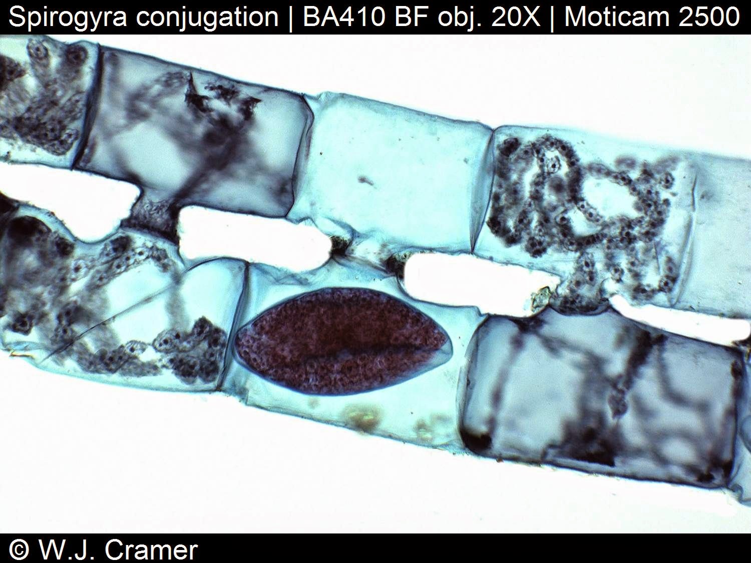
Conjugation in Spirogyra: the filaments show the typical results of scalariform ('ladderlike') conjugation. Two filaments lie parallel, and outgrowths from each filament grow towards each other, and then fuse to form a conjugation tube. The conjugation tubes allow the contents of one cell to transfer to the other. The dark oval shapes are diploid zygospores, formed after nuclear fusion between the two haploid cells. They form in only one of the two filaments. Sometimes more than two filaments take part in conjugation like a 'menage a trois'.
Spirogyra is a filamentous green alga in which the chloroplast has a characteristic spiral shape. In one of the photographs, you can see the chloroplast coiling against the outer edge of the cells. The numerous small round blobs along the edges of the chloroplast are the pyrenoids. The larger, faint blobs (they look like out-of-focus regions) that take up most of the volume of the cells in the filaments on the right are the nuclei, which are suspended in the interior of the cells.

Pyrenoids occur in many of the algae and are associated with the chloroplasts. Some of them are known to contain Rubisco, the enzyme that catalyzes the incorporation of inorganic CO2 into carbohydrate (Graham and Wilcox, 2000). In these algae, pyrenoids probably function to fix carbon. In other algae, pyrenoids are the sites of carbohydrate (typically starch) storage. Starch and iodine react to produce a deep blue- black color, so staining a thin algal prep with iodine will indicate the presence of pyrenoids.
Sources: Algalweb, MadSci Network
Conjugation in Spirogyra: the filaments show the typical results of scalariform ('ladderlike') conjugation. Two filaments lie parallel, and outgrowths from each filament grow towards each other, and then fuse to form a conjugation tube. The conjugation tubes allow the contents of one cell to transfer to the other. The dark oval shapes are diploid zygospores, formed after nuclear fusion between the two haploid cells. They form in only one of the two filaments. Sometimes more than two filaments take part in conjugation like a 'menage a trois'.
Spirogyra is a filamentous green alga in which the chloroplast has a characteristic spiral shape. In one of the photographs, you can see the chloroplast coiling against the outer edge of the cells. The numerous small round blobs along the edges of the chloroplast are the pyrenoids. The larger, faint blobs (they look like out-of-focus regions) that take up most of the volume of the cells in the filaments on the right are the nuclei, which are suspended in the interior of the cells.
Pyrenoids occur in many of the algae and are associated with the chloroplasts. Some of them are known to contain Rubisco, the enzyme that catalyzes the incorporation of inorganic CO2 into carbohydrate (Graham and Wilcox, 2000). In these algae, pyrenoids probably function to fix carbon. In other algae, pyrenoids are the sites of carbohydrate (typically starch) storage. Starch and iodine react to produce a deep blue- black color, so staining a thin algal prep with iodine will indicate the presence of pyrenoids.
Sources: Algalweb, MadSci Network
Tuesday, 29 July 2014
Do you like macro photography?
Do you like macro photography?
The macro lens included with any Moticam gives you endless opportunities…
Have a closer look at this sunflower using a Moticam 5 and a 12mm macro lens at different focus distances. You can see that the sunflower is in fact a composition of hundreds of individual flowers at different developmental stages.


Subscribe to:
Posts (Atom)