

Wednesday, 24 August 2016
A bio-indicator for air pollution
Growth of lichens on trees: Bio-indicator for clean and contaminated air. Lichens are dual beings, developed from a symbiosis of algae and tiny fungi. Fungi and algae alone are dependent on moisture.


Wednesday, 10 August 2016
Rock from the depths of the earth
Basalt belongs to the group of igneous rocks. Igneous rocks are formed when hot liquid magma (lava) from the depths of the earth, is forced to the surface by volcanic forces and flows like a mudslide directly over the earth's surface, where it cools down and solidifies. Basalt is used in the road and hydraulic engineering in particular, as a result of its favorable mechanical properties.

Basalt as (geological) young volcanic rock, gets a fine-grained structure when cooling down relatively rapid. It is interspersed with small circular hollow vesicles. The color ranges from dark gray via gray-black to dark blue. Basalt is very solid and

Basalt as (geological) young volcanic rock, gets a fine-grained structure when cooling down relatively rapid. It is interspersed with small circular hollow vesicles. The color ranges from dark gray via gray-black to dark blue. Basalt is very solid and
Monday, 1 August 2016
Introduction to Darkfield illumination - Slider solution
For transparent samples in light microscopy, dark field illumination is a simple and affordable contrast method. The idea of this technique is to display and to emphasize border structures, means abrupt changes of the refractive index within the sample. Especially for single-celled organism in fresh or sweet water environments, with a refractive index close to water, this contrast method gives aesthetic and informative images.
Dark field emphasizes borders and isolated, single structures, flagella of protozoa and tiny particles. The comparison of a diatom sample in bright field and dark field clearly shows this:

How such an image is achieved? We need to eliminate the direct light which ordinarily passes the specimen and which is responsible for the bright background in bright field illumination. For objectives with a Numerical Aperture ≤ 0.65 this
Dark field emphasizes borders and isolated, single structures, flagella of protozoa and tiny particles. The comparison of a diatom sample in bright field and dark field clearly shows this:

How such an image is achieved? We need to eliminate the direct light which ordinarily passes the specimen and which is responsible for the bright background in bright field illumination. For objectives with a Numerical Aperture ≤ 0.65 this
Wednesday, 27 July 2016
Tiny packages that contain all information necessary for life

Monday, 18 July 2016
The microscope objective - the key issue for best image performance
Different samples require different microscopes. This rule refers to the fact that an opaque, bulky sample with a reflective surface needs another treatment than a transparent, unstained smear from the cavitas oris. The microscope stand offers the necessary space for a correct positioning of the sample and all options for the appropriate illumination method.
The microscope objective is an even more specific item. Here we talk about the required resolution power (= numerical aperture), but also about cover glass correction, immersion method and working distance.
1. The standard upright microscope for transmitted light is constructed for glass slides with a 0.17mm cover slip. This restriction is indicated on the objective sleeve:
The cover slip has to be placed on top of the sample. A slight pressing of a dissecting needle will help to avoid too much embedding medium (water, etc.) between sample and cover slip. The embedding medium in this case works as an
The microscope objective is an even more specific item. Here we talk about the required resolution power (= numerical aperture), but also about cover glass correction, immersion method and working distance.
1. The standard upright microscope for transmitted light is constructed for glass slides with a 0.17mm cover slip. This restriction is indicated on the objective sleeve:
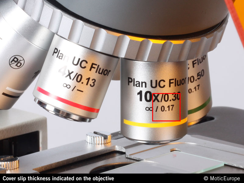 |
| Cover slip thickness indicated on Motic's Plan UC Fluor objectives |
The cover slip has to be placed on top of the sample. A slight pressing of a dissecting needle will help to avoid too much embedding medium (water, etc.) between sample and cover slip. The embedding medium in this case works as an
Thursday, 14 July 2016
Tar spot (Rhytisma acerinum)
The microscopic image shows an infected leaf with sclerotia. A consequence of monoculture.

Just like buildup of mold on pine needles and on the leaves of the willow, an ascomycete (Rhytisma) is the cause of tar spot. From late summer to autumn, round black mold deposits are growing on the leaves of several species of maple, which overwinter on the ground after the falling of the leaves. In spring distribution

Tuesday, 5 July 2016
When you still need a microscope to see Giant structures

Giant chromosomes can be observed in the salivary glands of certain two winged flies (Diptera) and were first observed by Balbiani in 1881 [1]. These chromosomes are oversized and develop from standard chromosomes when specialized cells
Wednesday, 29 June 2016
Rheinberg illumination

An interesting variance of darkfield is Rheinberg
illumination, discovered in 1896 in London by Julius Rheinberg. The major
difference between darkfield and Rheinberg illumination is color. Whereas in
darkfield, the background is black and the subject is white, Rheinberg goes a
step further and creates a colored background and a colored subject.
Monday, 27 June 2016
MOTIC’s New Tablet Solutions: enabling computer-free digital microscopy
Tablets and handheld computer devices are common place in every corner of working life. In schools, clinics or industrial applications, data gets captured, processed and transmitted using Android, Windows or iOS tablet devices.

Bringing this technology and flexibility to proper use in microscopy, Motic’s new Moticam Tablet solutions enable the user to choose the perfect solution for their circumstances. Following on the footsteps of Motic’s recent products to enable computer-free digital microscopy, the Moticam BTU8 and BTU10 feature a fully

Bringing this technology and flexibility to proper use in microscopy, Motic’s new Moticam Tablet solutions enable the user to choose the perfect solution for their circumstances. Following on the footsteps of Motic’s recent products to enable computer-free digital microscopy, the Moticam BTU8 and BTU10 feature a fully
Monday, 20 June 2016
The Dutch live with water
 |
| Micrasterias Rotata and Papillifera | Motic AE31E inverted Microscope | Moticam HD stack |
The Dutch have to live with water, because of their country lying below sea level for the greatest part. What does this have to do with microscopy?
Every year the Dutch are spending billions of euros in dikes, canals, rivers, bridges, locks, waste water purification, water quality monitoring, water research and innovation. Dutch water know how is being applied all over the world.

| Micrasterias Papillifera | Motic BA410E upright Microscope | Moticam 10 |
Subscribe to:
Posts (Atom)