- This plant is pollinated by bats.
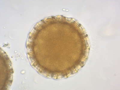
- Tthe large (~ 160 microns Ø) pollen grains of Cobaea show a complex structure which is worth to discover.
Pollen Grains in Brighfield
To give a full description, we should reactivate our Greek and Latin lessons from school:
Panto (from Greek: pan) = all, all over. And aperture (from Latin: apertura) = opening.
Cobaea pollen belongs to the “panto-aperturate” pollen class with openings for the pollen tube spread all over its surface. Further, the outer “skin”, the “exine”, shows a reticulate structure based on small rods. The pollen surface looks a little bit like a fine- patterned football.
To discover the full beauty of this pollen, we have to perform the “acetolysis”, a chemical treatment using acetic anhydride and sulfuric acid to increase the transparency of the pollen grains. For further details please refer to:

"Leitfaden der Pollenbestimmung:
für Mitteleuropa und angrenzende Gebiete"
Hans-Jürgen Beug
ISBN: 978-3899370430
"Pollen analysis"
Peter D. Moore
ISBN: 978-0865428959
©https://www.paldat.org/search/genus/Cobaea
The “inner surface” of the pollen grain shows the predefined apertures for the germination of the pollen tubes, while the optical “cross section” of the pollen grain displays the rods in longitudinal view.
Predefined pores for pollen tube germination
Rods in longitudinal section
Thanks to Klaus Laumeier, Cologne, Germany for the preparation of recent material


No comments:
Post a Comment