The “Escalón” cave, located in the Asón Valley in Cantabria (Spain), offers great possibilities for carrying out astrobiology work, and the company Astroland Agency has installed an operations base there, the “Ares Station”. From it, the sampling and recognition of biofilms have been organized, these grow on the humid walls of the cavity, in which these facilities are housed.
From the "Ares Station" different protocols have been tested to carry out research related to the study of the biofilms that cover the limestone rocks around the cave, which in this case, and unlike what happens in other cavities, are constituted, fundamentally, by cyanobacterial communities with species that are currently being investigated, and that are of great interest, because exceptionally scarce organisms have been found in them, and in some cases, not yet described.
Fig.1. The appearance of the cyanobacterial masses where this amoeba, Centropyxis sp. (aerophyla group) in the surroundings of the Ares station. Gloeobacter violaceus and Schizothrix sp. are some of the cyanobacteria among which it seems to be found most frequently.
These biofilms in the environment of the Ares Station have been revealed as a treasure of microbial biodiversity, and although most are made up of a complex and varied tapestry of cyanobacteria, they are also home to other microorganisms of great interest. Among them, some testate amoebas such as Difflugia alhadiqa, were discovered and described for the first time by our colleagues Carmen Soler-Zamora, Miguel González-Miguéns, and Enrique Lara, very recently, in the Hundidero cave, Montejaque, (Málaga, Spain). Another microorganism - that we show here and will have to be investigated more deeply - corresponds to a complex of amoebas of the genus Centropyxis, very close to the group of Centropyxis cassis, which present an enormous taxonomic difficulty.
Fig. 2. Naiara and Irene are high school students and they participate in this research work by assembling and preliminary observing a biofilm sample using equipment provided by Motic, the SMZ-168-TLED stereomicroscope.
The genus Centropyxis described by Stein in 1857 includes testate amoebas with a more or less discoidal and slightly flattened shell, with a flat or concave ventral side and a convex dorsal side. One of the most remarkable characteristics of this genre is that the opening of the shell, of very varied contours, is displaced towards one end.
A group of these fundamentally aquatic amoebas may have spines in a very varied number on their periphery, but this is not the case at hand. The species we are referring to here does not present them, although, like any of the representatives of the genus, its cover is covered with mineral particles, cemented by an organic matrix.
Fig.3. Centropyxis sp.(aerophyla group) among the cyanobacterial biofilm with Aphanothece saxicola dominance. Photographs taken at 400x magnification with the epifluorescence technique, with the FLED module and the Motic 40X/0.65/S (WD 0.6mm) dry objective. Equipment used: Motic Panthera CC trinocular.
More than 135 species and many varieties have been described, but many descriptions are currently being revised. Morphological recognition is insufficient to be able to accurately determine species with precision, which is only possible using genetic sequencing techniques.
There is currently an interesting debate about the identification of the species of the Centropyxis cassis complex, Centropyxis aerophila and Centropyxis constricta, which it is not appropriate to comment on in this publication, but it does seem clear that despite the fact that the morphology of our species is closer to Centropyxis cassis due to the shape of the theca, its dimensions are far from being those of the latter, which in C. cassis oscillate between 54×60 µm wide and 64×75 µm long. In the few specimens found among the biofilm of the cave, the dimensions range between 35–40 µm wide and 50–58 µm long and 20–15 µm in diameter, in the major and minor axes, respectively.
The form of C. constricta is very reminiscent of that of the species discussed in this article, but its size is much larger (about 100 µm) and its habitat is completely different - it lives in bright and cool areas with Sphagnum.
Fig.4. Centropyxis sp.(aerophyla group) in another view among the cyanobacterial biofilm with a domain of Aphanothece saxicola. Photographs taken at 400x magnification with the epifluorescence technique, with the FLED module and the Motic 40X/0.65/S (WD 0.6mm) dry objective. Equipment used: Motic Panthera CC trinocular.
From C.aerophyla, with almost spherical contours and larger dimensions, it seems to be further away both because of its shape and size and because of its habitat.
Everything seems to indicate that the species we show here is different and that it could be a new species. All this will have to be confirmed in subsequent sampling and research, in which the sequencing of its genetic material will be key.
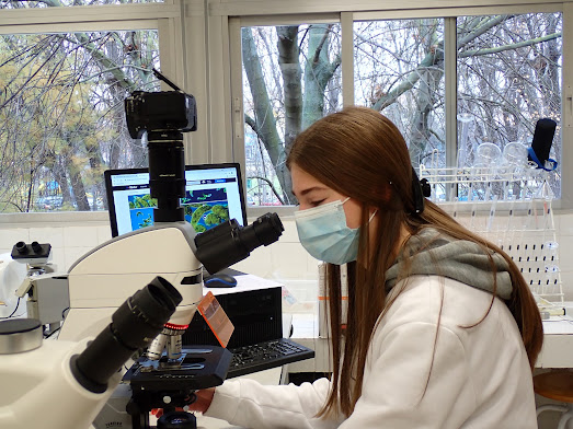
Fig.5. Observing the samples of Centropyxis sp. (aerophyla group) with the FLED module and the Motic 40X / 0.65 / S (WD 0.6mm) dry objective. Students of 1º of Baccalaureate of the subject Scientific Culture in the “IES Escultor Daniel" (Logroño, Spain).
Possibly Enrique Lara's team from the “Real Jardín Botánico” of Madrid and other experts such as Edward Mitchell or Foissner and Korganova, who have worked on this genre, can make their contributions and comments.
In any case, it is a relevant finding and further proof of the interest presented by these barely studied biofilms.
All the photographs have been taken at a magnification of 400, with bright field and epifluorescence techniques, with a Motic Panthera CC trinocular equipment and come from the samples collected inside the Escalón cave, in the Ares Station environment. It is a very dimly lit area where the Astroland Agency is developing an approach to learning about Mars in its astrobiological project.
Fig.6. Centropyxis sp.(aerophyla group) from a fresh sample of the cyanobacterial biofilm. Photographs taken at 400x magnification with the epifluorescence technique, with the FLED module and the Motic 40X/0.65/S (WD 0.6mm) dry objective. The opening of the theca and the contour of this amoeba can be seen very well, as well as some cells of the Gloebacter colonies in yellow. Equipment used: Motic Panthera CC trinocular.
Today this amoeba sees the light from the darkness of the cave where it was found for those who come to read this publication and for the students of the sculptor Daniel High high school in Logroño. They have participated in these observations handling the Motic equipment - both microscopes and stereomicroscopes - learning how to handle this equipment both with conventional brightfield lighting and with the FLED epifluorescence module, essential for identifying and seeing the mineral particles that make up the theca of this amoeba.


No comments:
Post a Comment