Monday, 28 February 2022

Rare Disease Day 2022

Today is the world day of rare diseases, an opportunity to raise awareness and act, to draw attention to the needs of a community that has over 300 million people in the world.

A disease or condition is rare if it affects fewer than 1 in 2.000, but thanks to the increase of its awareness now #RareIsMany. 

This increase is the result of changes in society, including the active promotion of rare diseases in general. Every person living with a rare disease is unique, but shared experiences are what unite.

Let’s keep inspiring and encouraging projects spanning all medical areas and contributing to understanding the causes and characteristics of rare diseases, developing new diagnoses and therapies for patients, and promoting best practices for use in hospitals and health systems.

Technology plays a significant role in the diagnosis of rare diseases: it allows for the pooling of resources, data, and complementary skills, which no single individual could obtain alone.

For medical technology that improves care, for equity in health care, for access to early diagnosis and treatment for people with a rare disease.

Fig. 1 MoticEasyScan: Auto-focus, overview, and detail information for effective acquisition of all necessary slide information. The FS-Live Telepathology System allows health specialists to view a case slide in seconds from anywhere in the world, improving the quality of care for patients. 
Fig. 2 https://www.rarediseaseday.org/

Bones, teeth, and minerals

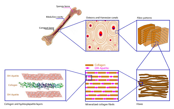
The endoskeleton of many animals is made up of rigid, solid, and resistant organs which are the bones. Bones, despite their inert appearance, are alive and contain cells, blood vessels, nerves, fat, bone marrow. They are in constant transformation. Bone tissue is the basis of its structure. Bone cells are only 2% of bone tissue, the rest is the extracellular matrix. This matrix is made up of 30% organic substances such as the protein called collagen. The remaining 70% is an inorganic substance: hydroxylapatite.

The solidity of the bone tissue is given by the presence of hydroxylapatite crystals that are arranged around the collagen fibres forming an important framework, giving rise to the exceptional mechanical properties of bones (figure 1). A fairly accurate comparison would be the structure of a building, where the collagen would correspond to the steel rods and the hydroxylapatite crystals to the concrete that surrounds them (UCM, online).

Figure 1- Bone structure. From the macroscopic to the nanometric and atomic world.
Scheme based on Nair et al. (2013) and Wang et al. (2017).

But it is not only in the bones that hydroxylapatite can be found. Although bones seem very strong, the hardest substance in our body is tooth enamel, which is also made up of hydroxylapatite.

As it can be seen, OH-apatite is very present in the organic world, the world of living beings. But it can also be found in nature, in the inorganic world, in the mineral kingdom.

But what is hydroxylapatite?

Within the classification of minerals is the apatite supergroup. Its nomenclature and classification was approved by the International Mineralogical Association in 2010 (Pasero et al.).

The members of this supergroup have a generic formula according to:

M1M2(TO)₃ X
Where:

T= P⁵⁺, As⁵⁺, V⁵⁺, Si⁴⁺, S⁶⁺, B³⁺

M1 and M2 are represented by numerous cations in the periodic table (in two different positions, 1 and 2) including the rare earth elements (REE): Ca²⁺, Pb²⁺, Ba²⁺, Sr²⁺, Mn²⁺, Na⁺, Ce³⁺, La³⁺, Y³⁺, Bi³⁺.

And to complete the formula, X would correspond to: (OH)⁻, F⁻ and/or Cl⁻.

Members of the apatite supergroup, about 50 species, show a wide range of solid solutions due to the substitution of the forming atoms. This supergroup is currently divided into five divisions: apatite group, hedyphane group, belovite group, britholite group, ellestadite group.

The one of interest in this note is the group of apatite, which includes several species between phosphates, arsenates, and vanadates, some well-known as pyromorphite, mimetite, or vanadinite, all of them from the hexagonal system (figures 2, 3, and 4).

Figure 2 - Mimetite crystal with cerussite and calcite.

Calcium phosphates are found in this group, which have a simplified formula according to Ca₅(PO₄)₃X

where X = F (fluorapatite), Cl (chlorapatite), OH (hydroxylapatite). 

They are often colloquially called "apatite". In nature, the most common are fluorapatite and hydroxylapatite.

Geologically, members of this supergroup are found in different types of igneous, sedimentary, and metamorphic rocks. Even in meteorites (ordinary chondrites L) or in the basaltic breach in Martian meteorites (impactites).

 “Apatite”: where does it come from?

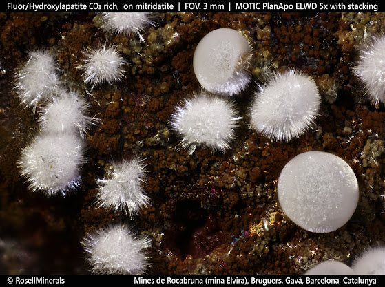
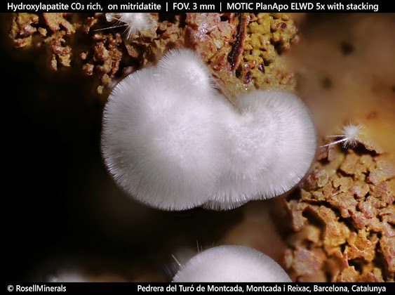
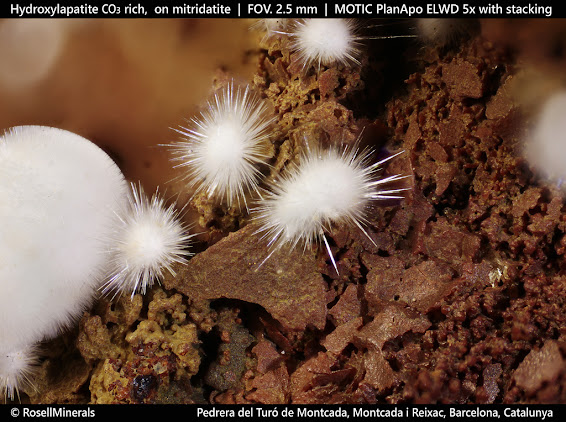
The specimens in the photographs correspond to two classic Catalan deposits: Turó de Montcada (Montcada i Reixac) and the Rocabruna mines (Bruguera, Gavà), both in the province of Barcelona. In Catalonia, there are other deposits with similar geology, more or less rich in phosphates, such as Torroja del Priorat, Cornudella de Montsant, Figuerola del Camp, Santa Creu d'Olorda or Pineda de Mar, among others. 

They are located mainly in the Catalan Coastal Ranges, which run parallel to the coast, from the south of the province of Tarragona to Girona.

These deposits of stratiform sedimentary origin share a similar origin and are found in materials from the Paleozoic series: shales, limestones, dolomites, and quartzites.

In these deposits, Silurian shales (about 420 million years BP) contain rich levels of marine sedimentary phosphates (phosphorites). When meteoric fluids pass through these shales with pyrite (FeS2), they become acidic (form sulphuric acid) and dissolve them. These fluids, rich in phosphorus, reach areas where carbonate rocks predominate, producing the neutralization of acidity and the precipitation of various minerals, including fluorapatite and hydroxylapatite.

Figure 3 - Divergent and globular aggregates of acicular crystals, on a matrix with goethite and mitridatite.

Figure 4 - Divergent aggregates of acicular crystals, on a matrix with goethite and mitridatite (RM3430).

Figure 5 - Divergent aggregate of acicular crystals, on a matrix with goethite and mitridatite (RM3430). 

It should be noted that many of the "apatite" formed by these processes incorporate in their structure carbonate anions (CO₃²⁻) which replace the phosphate anions (PO₄³⁻).
It follows that the varieties carbonate-rich fluorapatite and carbonate-rich hydroxylapatite are present: Ca₅(PO₄,CO₃)₃(F, OH).

This is the case with the specimens in figures 3, 4, and 5.

Figure 6 - SEM image of the specimen from the Turó de Montcada (RM3430).

Differentiating whether it is fluorapatite or hydroxylapatite requires a chemical analysis, with scanning electron microscopy being an ally that is sometimes not very sensitive. In the specimens studied in these two Catalan mines, the presence of both species has been confirmed, but distinguishing those that have fluorine from those that contain hydroxyl groups is not an easy task and impossible with the naked eye (figures 6 and 7).

Figure 7 - SEM-EDS of the specimen from Turó de Montcada (RM3430). The presence of fluorine is not observed in any of the studied points of this specimen. The hexagonal shape of the needles can be observed. 

Nature uses the elements at its disposal to form both organic and inorganic structures, mixing both worlds. In this respect, human beings are part of both.


References

Nair, A.K., Gautieri, A., Chang, S.W., Buehler, M.J. (2013): “Molecular mechanics of mineralized collagen fibrils in bone”. Nature Commun. 4, 1724.

Pasero, M., Kampf, A.R., Ferraris, C., Pekov, I.V., Rakovan, J., White, T.J. (2010): “Nomenclature of the apatite supergroup minerals”. European Journal of Mineralogy, 22, 163-179. https://www.researchgate.net/publication/228477092 [on-line 22 Febrero 2022].

Wang, W., Yeung, K.W.K. (2017): “Bone grafts and biomaterials substitutes for bone defect repair: A review”. Bioactive Materials, 2, 224-247. https://www.researchgate.net/publication/317392938 [on-line 22 febr. 2022].

[web] Fisiopatología ósea. Universidad Complutense de Madrid. https://www.ucm.es/data/cont/docs/420-2014-02-18-01%20fisiopatologia%20osea.pdf [on-line 22 febrero 2022].


Friday, 25 February 2022

Trametes versicolor, turkey tail

Turkey tail (Trametes versicolor a bracket fungus species) is a species of fungus that closely resembles a turkey tail. It forms stacked terraces with a wavy white growth edge. Within this edge, areas are having a variety of colors such as white, black, beige, fir, gray, brown, and blue. That is why it is named ‘versicolor’. The terraces have a size of up to 8 centimeters and are only a few millimeters thick. As a polypore, turkeys tail holds its spores in white or yellowish tubes, so its underside displays tiny holes visible to the naked eye. The spores are white to pale yellow. The mushroom meat is tough and not edible. The fungus causes white rot in wood, breaking down either the lignin or cellulose.

Fig 1. Turkey tail top 

Fig 2. Turkey tail underside

Turkey tails are possibly the most common mushroom you will find. Their uses are mainly medicinal and decorative. They can have great beauty. They dry easily to become leathery tough. The material used for the microscopic image has been stained for better visibility.

Fig 3. Trametes versicolor c.s. analyzed with a SWIFT 3-B, 10X objective microscope and a Moticam 5
© www.willemsmicroscope.com

Thursday, 24 February 2022

The passion for educational microscopy

It is sometimes the actions that become a lasting memory. As an ambassador of the necessity of science for the continuity of life, Marylène Hermouet has been to schools and universities. She dedicated her life to this very task. 

That is what she stood for, that is what she fought for.

Education is the future of science and Marylène strongly believed in it. 

To advance in the educational and scientific fields interdisciplinary it is necessary to cultivate a research-based approach to develop the practice of young students.

Marylène knew that promoting critical thinking and analytical skills through hands-on learning helps define both academic and personal interests.

The microscope that Marylène used to show the application of technology in an educational space, allowing the use of more interactive tools, has been given to the Pathological Anatomy Department of the Sant Joan de Deu Hospital in Barcelona (Spain), to be a useful tool for research and prevention of new and passionate scientists, as a profound recognition of Marylène. 


In the picture is the BA410 Elite microscope by Maryléne Hermouet in its new location at the Sant Joan De Deu hospital, between the Head of the Pathological Anatomy Service, Cristina Jou Muñoz, and Emilie Blasco, France Sales Manager at Motic Europe.

Wednesday, 23 February 2022

We have an army in our bodies!

They are usually simplified as “white cells”, come from different places, have different functions, names…  and help us protect ourselves from antigens. That is, in a nutshell, what we can pretty much expect from them.

Sometimes, as we know, some cells escape the normal mechanisms of our bodies. They became immortal, rapidly reproducing themselves, and go to places not supposed to be (metastasis). That’s what we call cancer.

Figure 1. Histiocytoma is a common tumor among canine skin diseases that occurs mainly on the head, ears, or limbs of animals under four years of age.

We should expect our “white cells” to protect us from cancer, but sometimes they are cancer. Have you ever heard about “blood cancer” or Leukaemia? 

In this miniseries, you’ll learn about some “white cells” cancer entities. 

Are you ready for it?

© www.willemsmicroscope.com

Thursday, 17 February 2022

Eating raw meat?

The Trichinella spiralis parasite is found in animals, especially those living in the wild. It can occur in raw pork, raw horse meat or raw meat from games such as wild boar.


Section of infected muscle with encysted larvae. Motic Panthera U with Plan Apo 40X objective and Moticam 10+.

The parasite is a microscopic worm whose larva moves through the body. Muscles, in particular, suffer from these larvae. 

The larva of the Trichinella spiralis can cause the disease trichinosis or trichinellosis in humans. The symptoms are abdominal pain, nausea, fever, and diarrhea in the first 2 weeks.

Over time, edema, itching, fever, and chronic muscle aches occur. In severe cases, weight loss, dehydration, heart, and respiratory problems, and brain inflammation can occur. A fatal outcome is rare in Western Europe but cannot be excluded. 

The severity of the disease depends on the number of parasites you have ingested. 

You can become infected if you eat raw meat or insufficiently heated meat. Heating above 70 ° C kills the parasite.

Prepared slide by Lieder www.lieder.com

© www.willemsmicroscope.com

Wednesday, 16 February 2022

Schizothrix calcicola: the filaments of fire

The biofilms that cover the walls of the “La Cueva del Escalón” cave, located in the Ason Valley (Cantabria, Spain), have proved to be a treasure trove of microbial biodiversity. They are mostly made up of a complex and varied tapestry of cyanobacteria, many of them little known, and which are currently being investigated in several projects.

The "Ares Station" is an operational center of the "Astroland Agency" company, installed inside the cave itself, from where part of the research work on biofilm takes place and basically acts as a support for sampling activities. These are environmentally friendly structures in which they are located, and from where an interesting astrobiology program is developed, in which different protocols related to microbiological research and the appearance of life on our planet are tested, or looking for signs of life on Mars.

Among the multicolored jellies that form the dense tapestry that covers the walls of the cave, those of the cyanobacterium Schizothrix are characterized by their green color and can appear by spraying irregularly other jelly-like masses of pink, grayish, and other various colors, or occur among other masses cyanobacteria showing fibrous structures.

Fig. 1. Macroscopic appearance of Schizothrix calcicola colonies, seen as green gelatinous masses among other filamentous cyanobacteria such as Scytonema julianum.

Under the microscope, using the Motic Panthera equipment, the filaments of Schizothrix calcicola, using objectives of different magnifications, reveal themselves as tenuous, narrow, and fragile structures, and appear to be formed by cells of just over a micron in width and four or five microns in diameter in length. One after another, these cells gently curve their long chains as they tangle.

Fig. 2. Aspects presented by filamentous colonies of Schizothrix calcicola when viewed with an epifluorescence microscope at 400X magnification and in a specimen that has been in a cold cell for 5 months in the dark. The photographs were taken with a Motic Panthera CC trinocular equipment with the Motic FLED fluorescence module. Image captured with a 40X / 0.65 / S (WD 0.6mm) objective. 

All these strands are wrapped in thick sheaths of transparent and fibrous gelatin, which end in a long tip and protect the Schizothrix groups by preventing them from drying out.

The filaments in Schizothrix calcicola are irregularly arranged to form a mucilaginous, unincorporated thallus; with 1 - 2 (3) trichomes per pod, parallel and sometimes slightly curved. Colorless pods closed apically and sharp. Trichomes can be 0.7 - 1.5 (2) μm in diameter, are pale bluish-green in color with cells longer than wide, (1) 2 - 4.4 μm in length, and terminate in an apical cell rounded.  

Fig.3. Schizothrix calcicola filaments photographed at 1000x magnification with the brightfield technique using the Motic 100X / 1.25 / S-Oil immersion objective (WD 0.16 mm).

All this tangle, which appears inert when viewed under brightfield illumination, turns out to be completely different when viewed illuminated with Motic's FLED fluorescence module and reveals its life by emitting a red-orange light that resembles the colors of a flame. in the form of filaments.

Fig. 4. Filaments of Schizothrix calcicola from a sample stored in cold storage and in the dark after 5 months. Images taken at 400x magnification with the epifluorescence technique, with the FLED module and the Motic 40X / 0.65 / S dry objective (WD 0.6mm). In the lower part can be seen the cells of some colonies of Gloeobacter violaceus colored with intense yellow color.

The name Schizothrix calcicola includes a complex of at least twenty morphological and genotypic varieties awaiting revision and which in Europe live on humid limestone rocks in temperate regions. In this cave of Escalón it is part of the flora that covers the wet walls in the areas near the entrance.

The images, taken at 400 and 1000 magnifications, with brightfield and epifluorescence techniques, with Motic Panthera CC trinocular equipment, come from samples collected inside the Escalón cave, near the Ares station, in a very poorly lit area in that the "Astroland Agency" is developing in its approximation project to the knowledge of Mars.

Fig. 5. Filaments of Schizothrix calcicola from a fresh specimen. Images taken at 400x magnification with the epifluorescence technique, with the FLED module and the Motic 40X / 0.65 / S dry objective (WD 0.6mm). Other cyanobacterial forms of different genera can be seen around the reddish filaments of Schizothrix calcicola, showing another coloration.

It is striking how these samples observed today and collected in late June 2021 continue to maintain vital and photosynthetic activity after being in a cold room for almost six months.

Fig. 6. Observation of samples of Schizothrix calcicola with the FLED module and the Motic 40X / 0.65 / S (WD 0.6mm) dry objective. Students of the subject Scientific culture at the high school "Escultor Daniel" (Spain)

Today these cyanobacteria are shown again to the readers of this publication and for the 1st year the students of the "Escultor Daniel"  high school in Logroño (Spain), who participated in these observations learning to handle the Motic equipment,  both with conventional brightfield illumination, as with the FLED epifluorescence module, essential for detecting the presence of photosynthetic activity in any sample of cyanobacteria.

Tuesday, 8 February 2022

The jelly amethyst: Gloeobacter Violaceus

The “Escalón Cave”, located in the Asón Valley (Cantabria) has been revealed as a treasure of microbial biodiversity, in which the biofilms that cover the walls of this cavity, show a complex and varied tapestry of cyanobacteria, many of them little known and that they are currently being investigated.

Image 1: General appearance of the biofilm near the entrance to the cave of the Escalón (Cantabria, Spain)

In this context, the space agency Astroland has installed an operations center, the "Ares Station”, inside the same cave. These facilities are respectful of the environment in which they are located, and from where part of their program of activities is carried out, including an interesting astrobiology program, in which different protocols related to microbiological research and the appearance of life on our planet, or the search for signs of life on Mars.

Image 2: The Ares Station 

The Ares Station is an operational work and research center that aims to recreate an environment analogous to that of Mars in the cavities that exist on this planet.

Some of the cyanobacteria that live as part of the biofilm that covers the walls of the cave, like Gloeobacter, are very interesting and can help to understand what photosynthetic organisms have been able to evolve, perhaps since those that the red planet had seas like those of the Earth and the first brushstrokes of life of colors, they were brushstrokes of gelatine, perhaps also violet and pink gelatine, like the one’s masses of these cyanobacteria investigated in this project, using for it the Motic Panthera microscope, equipped with an epifluorescence illumination module.

Image 3: General appearance of Gloeobacter violaceus nearby of the "Ares Station"

The cyanobacterium Gloeobacter violaceus has one of the most complex photosynthetic apparatus primitives that is known, probably the ancestor of all the photosynthetic apparatus of all the green organisms that inhabit our planet today.

Gloeobacter violaceus thus recalls, sheltered in a humid and gloomy cave, the time of an origin in which the world was uninhabitable, and in which forms like it took the first steps opening the path of life.

It is a living and unique treasure that grows sheltered from the wind, pink or violet-like an amethyst when it grows in mass.

Internally it shows a unique ancestral cell organization, an uncommon structure of the photosynthetic apparatus characterized by a complete absence of internal membranes (thylakoids). The phylogenetic investigations that have been carried out to date with this organism, whose genetic material has been completely sequenced, demonstrate its basal position among all organisms with organelles capable of photosynthesis.

Image 4: Observation of Gloeobacter samples with the Motic Panthera microscope equipped with an epifluorescence module. Student of the "Scientific Culture" course at a high school in Logroño (Spain).

"Gloeobacter violaceus" has become one of the key species in the evolutionary study of photosynthetic life. It is also among the most widely used organisms in experimental photosynthesis research.

The individuals of "Gloeobacter violaceus" are formed by solitary cells or aggregated in irregular groups, broadly oval or rod-shaped, and surrounded by delimited and narrow gelatinous envelopes, which contain one or more cells, and whose ends generally present a granular structure. Cells are not mobile and cell division is carried out by simple binary transverse fission, perpendicular to the long axis of the cell. Reproduction is probably by the release of single enveloped cells or small groups of cells.

Image 5: General appearance of a Gloebacer specimen mounted in vivo and observed at x400 magnification with the brightfield technique with the Motic Panthera equipment.

Image 6: The same sample as above and the same field of view imaged using the epifluorescence module. The contours of the Gloebacter cells stand out in yellow due to the excitation of the chlorophylls they contain.

The forms that give rise to the purple gelatines are wider and present a stratified mucilaginous envelope in several layers, these, which we photograph here today, with the Motic Panthera microscope, present a much more elongated and narrow body and are only enveloped in a gelatine layer, barely perceptible and not stratified.

Image 7: General appearance of a Gloeobacter sample mounted in vivo and observed at magnification x1000 with the epifluorescence technique with the Motic Panthera equipment. The tenuous mucilaginous envelopes that surround some of the cells can be appreciated.

It is unknown if these elongated forms of the pink jellies correspond to a phase of the life cycle of those that grow in amethyst jellies or if it is a genetic variant of them. In any case, their biological interest is undeniable due to the extraordinary efficiency with which they can carry out photosynthesis practically in the absence of light for our eyes.

It is striking how these samples observed today and collected at the end of June 2021 continue to maintain vital and photosynthetic activity after remaining in a cold room for almost six months.

Gloeobacter violaceus is a treasure. From "Proyecto Agua" - a scientific and informative project that tries to make known the world of the microscopic through a Flickr gallery (https://www.flickr.com/photos/microagua/) it has been found and documented for the first time in the Iberian territory.

It is a species that is hardly known until now from any other locality in the Swiss Alps and Mexico. It is truly a unique soft living fossil, valuable as the most valuable of treasures to understand how photosynthetic organisms began to take the first steps on our planet and made life possible for others, including ours.

Image 8- first photographs of Gloeobacter violaceus taken in Iberian territory from Proyecto Agua with DIC at 400x magnification.

Gloeobacter violaceus lives in conditions of very little light and high humidity on the limestone walls of the caves, on which it forms a film of irregular thickness, gelatinous consistency, and varied color that ranges between pink, violet, and amethyst. With its forms of light in its soft galaxies, it transports us to the past, to the lost times in which the life of the Planet began to shine as these cyanobacteria do today, a soft jewel of amethyst.

The images taken with the Motic Panthera equipment with the x40 and x100 lenses and the epifluorescence module (Images 4, 6, and 7) allow us to perfectly distinguish the contours of these cyanobacteria that emit an intense yellow fluorescence, which is very characteristic. It is striking how, with the bright field technique taken with the same equipment and the same objectives (Image 5), these organisms are practically unrecognizable in the gelatinous mass in which they live.

Friday, 4 February 2022

World Cancer Day 2022

Cancer is still one of the most common causes of death in the world today. On World Cancer Day, we express our appreciation and admiration to all survivors, research fighters, and cancer patients.

Although today is World Cancer Day, the persistence of scientific research for years has added small steps towards healing. It is precisely these small steps and tangible results that allow new approaches to early diagnosis, giving hope because they alleviate struggle and protect life.

Again and again, research has managed to develop new treatment methods. According to research results, the mRNA vaccines were used in the treatment of cancer. Research has been carried out on how mRNA technology can be used in cancer treatment for about 30 years. This treatment is intended to detect and destroy malignant immune cells. However, there are still some hurdles to overcome, as it is much more difficult to develop an mRNA vaccine against endogenous cancer than a vaccine against an external attacker such as the coronavirus.

MoticEasyScan Pro 6 – DS Server software 

The diagnosis of cancer in its earliest stages often offers the best chance of cure. It is very important to ensure early diagnosis and detection.

Prevention is the best weapon to overcome cancer, and it is within our reach every day: follow a balanced and healthy diet, exercise and undergo regular medical check-ups.