Tuesday, 8 February 2022

The jelly amethyst: Gloeobacter Violaceus

The “Escalón Cave”, located in the Asón Valley (Cantabria) has been revealed as a treasure of microbial biodiversity, in which the biofilms that cover the walls of this cavity, show a complex and varied tapestry of cyanobacteria, many of them little known and that they are currently being investigated.

Image 1: General appearance of the biofilm near the entrance to the cave of the Escalón (Cantabria, Spain)

In this context, the space agency Astroland has installed an operations center, the "Ares Station”, inside the same cave. These facilities are respectful of the environment in which they are located, and from where part of their program of activities is carried out, including an interesting astrobiology program, in which different protocols related to microbiological research and the appearance of life on our planet, or the search for signs of life on Mars.

Image 2: The Ares Station 

The Ares Station is an operational work and research center that aims to recreate an environment analogous to that of Mars in the cavities that exist on this planet.

Some of the cyanobacteria that live as part of the biofilm that covers the walls of the cave, like Gloeobacter, are very interesting and can help to understand what photosynthetic organisms have been able to evolve, perhaps since those that the red planet had seas like those of the Earth and the first brushstrokes of life of colors, they were brushstrokes of gelatine, perhaps also violet and pink gelatine, like the one’s masses of these cyanobacteria investigated in this project, using for it the Motic Panthera microscope, equipped with an epifluorescence illumination module.

Image 3: General appearance of Gloeobacter violaceus nearby of the "Ares Station"

The cyanobacterium Gloeobacter violaceus has one of the most complex photosynthetic apparatus primitives that is known, probably the ancestor of all the photosynthetic apparatus of all the green organisms that inhabit our planet today.

Gloeobacter violaceus thus recalls, sheltered in a humid and gloomy cave, the time of an origin in which the world was uninhabitable, and in which forms like it took the first steps opening the path of life.

It is a living and unique treasure that grows sheltered from the wind, pink or violet-like an amethyst when it grows in mass.

Internally it shows a unique ancestral cell organization, an uncommon structure of the photosynthetic apparatus characterized by a complete absence of internal membranes (thylakoids). The phylogenetic investigations that have been carried out to date with this organism, whose genetic material has been completely sequenced, demonstrate its basal position among all organisms with organelles capable of photosynthesis.

Image 4: Observation of Gloeobacter samples with the Motic Panthera microscope equipped with an epifluorescence module. Student of the "Scientific Culture" course at a high school in Logroño (Spain).

"Gloeobacter violaceus" has become one of the key species in the evolutionary study of photosynthetic life. It is also among the most widely used organisms in experimental photosynthesis research.

The individuals of "Gloeobacter violaceus" are formed by solitary cells or aggregated in irregular groups, broadly oval or rod-shaped, and surrounded by delimited and narrow gelatinous envelopes, which contain one or more cells, and whose ends generally present a granular structure. Cells are not mobile and cell division is carried out by simple binary transverse fission, perpendicular to the long axis of the cell. Reproduction is probably by the release of single enveloped cells or small groups of cells.

Image 5: General appearance of a Gloebacer specimen mounted in vivo and observed at x400 magnification with the brightfield technique with the Motic Panthera equipment.

Image 6: The same sample as above and the same field of view imaged using the epifluorescence module. The contours of the Gloebacter cells stand out in yellow due to the excitation of the chlorophylls they contain.

The forms that give rise to the purple gelatines are wider and present a stratified mucilaginous envelope in several layers, these, which we photograph here today, with the Motic Panthera microscope, present a much more elongated and narrow body and are only enveloped in a gelatine layer, barely perceptible and not stratified.

Image 7: General appearance of a Gloeobacter sample mounted in vivo and observed at magnification x1000 with the epifluorescence technique with the Motic Panthera equipment. The tenuous mucilaginous envelopes that surround some of the cells can be appreciated.

It is unknown if these elongated forms of the pink jellies correspond to a phase of the life cycle of those that grow in amethyst jellies or if it is a genetic variant of them. In any case, their biological interest is undeniable due to the extraordinary efficiency with which they can carry out photosynthesis practically in the absence of light for our eyes.

It is striking how these samples observed today and collected at the end of June 2021 continue to maintain vital and photosynthetic activity after remaining in a cold room for almost six months.

Gloeobacter violaceus is a treasure. From "Proyecto Agua" - a scientific and informative project that tries to make known the world of the microscopic through a Flickr gallery (https://www.flickr.com/photos/microagua/) it has been found and documented for the first time in the Iberian territory.

It is a species that is hardly known until now from any other locality in the Swiss Alps and Mexico. It is truly a unique soft living fossil, valuable as the most valuable of treasures to understand how photosynthetic organisms began to take the first steps on our planet and made life possible for others, including ours.

Image 8- first photographs of Gloeobacter violaceus taken in Iberian territory from Proyecto Agua with DIC at 400x magnification.

Gloeobacter violaceus lives in conditions of very little light and high humidity on the limestone walls of the caves, on which it forms a film of irregular thickness, gelatinous consistency, and varied color that ranges between pink, violet, and amethyst. With its forms of light in its soft galaxies, it transports us to the past, to the lost times in which the life of the Planet began to shine as these cyanobacteria do today, a soft jewel of amethyst.

The images taken with the Motic Panthera equipment with the x40 and x100 lenses and the epifluorescence module (Images 4, 6, and 7) allow us to perfectly distinguish the contours of these cyanobacteria that emit an intense yellow fluorescence, which is very characteristic. It is striking how, with the bright field technique taken with the same equipment and the same objectives (Image 5), these organisms are practically unrecognizable in the gelatinous mass in which they live.

Friday, 4 February 2022

World Cancer Day 2022

Cancer is still one of the most common causes of death in the world today. On World Cancer Day, we express our appreciation and admiration to all survivors, research fighters, and cancer patients.

Although today is World Cancer Day, the persistence of scientific research for years has added small steps towards healing. It is precisely these small steps and tangible results that allow new approaches to early diagnosis, giving hope because they alleviate struggle and protect life.

Again and again, research has managed to develop new treatment methods. According to research results, the mRNA vaccines were used in the treatment of cancer. Research has been carried out on how mRNA technology can be used in cancer treatment for about 30 years. This treatment is intended to detect and destroy malignant immune cells. However, there are still some hurdles to overcome, as it is much more difficult to develop an mRNA vaccine against endogenous cancer than a vaccine against an external attacker such as the coronavirus.

MoticEasyScan Pro 6 – DS Server software 

The diagnosis of cancer in its earliest stages often offers the best chance of cure. It is very important to ensure early diagnosis and detection.

Prevention is the best weapon to overcome cancer, and it is within our reach every day: follow a balanced and healthy diet, exercise and undergo regular medical check-ups.

Wednesday, 26 January 2022

Pericarditis

Pericarditis is an inflammation of the pericardium. This is a solid double membrane around the heart. The innermost layer is fused with the heart muscle. The pericardium protects the heart from external influences and keeps the heart in place. Between the two layers of the pericardium, there is a small amount of clear liquid, which makes it easy for the layers to slide past each other (like two glass plates slide easily over each other with water in between).

In acute pericarditis, complaints such as severe pain in the chest area can occur in the beginning. The pericardium contains many nerves. The friction between the membranes of the inflamed pericardium causes severe pain. The pain increases when you move, breathe in, or lie flat.

The causes of pericarditis are:

• virus infection (for example after flu or cold)
• open heart surgery
• heart attack
• bacterial infection (rare)
• other illnesses

Sometimes the cause remains unknown.

In the pictures, we see part of the heart muscle tissue (myocardium) on the left, with adipose (fat) tissue to the right. On its right side, numerous leukocytes, infiltrated as a result of the inflammation are visible.

Pericarditis, heart c.s. - PlanApo 4X 

Pericarditis, heart c.s. - PlanApo 20X

Pericarditis, heart c.s. - PlanApo 40X

Note: the prepared slide, a collector’s item, is 60 years old.

With thanks to Herbert Spoon, Doctor of Medicine.

© www.willemsmicroscope.com

Monday, 24 January 2022

Metazeunerite-kahlerite from the Atrevida Mine, Vimbodí i Poblet, Conca de Barberà, Catalunya

The presence of uranium minerals in the Atrevida mine has been known for years. The first notice was published in Mineralogistes de Catalunya (Viñals et al., 1990; Bareche, 1990). A posteriori, the presence of uranium in the Silurian and Carboniferous deposits in the Prades mountains area was described by Carles Canet (Canet, 2001).

The article of Bareche states that uranium minerals were collected in an open pit with a huge size baryte vein. It is also reported that Geiger counter research throughout the demarcation showed no signs of other uranium mineralizations. However, in the Tacha mine there are certain materials (with baryte and nickel minerals) that show a certain radioactive emission.

Crystals identified as kahlerite-zeunerite. Col. E. Bareche (image available in the Joan Astor photos
Museu Mollfulleda de Mineralogia d’Arenys de Mar and online: Issuu, Astor, 2014).

Figure 2.- Zeunerite crystals. Col. E. Bareche. Photo: Joan Astor (image available in the Joan Astor photos
Museu Mollfulleda de Mineralogia d’Arenys de Mar and online: issuu, Astor, 2014).

In the image that illustrates the article (figure 1) it can be seen a group of apple-green tabular crystals, pseudohexagonal, with small brown globular aggregates, identified as kahlerite-zeunerite. To reach this conclusion an EDS spectrum is presented where can be seen some elements: arsenic, uranium, copper, and iron.


Figure 3.- EDS spectrum of the specimen in Figure 1 (Bareche, 1990).

Kahlerite is a hydrated uranyl and iron arsenate of the chemical formula Fe2+(UO2)2(AsO4)2·12H2O, tetragonal crystalline system, belonging to the Autunite Group. It was discovered in 1953 and is named in honour of Franz Kahler (1900-1995), a geologist at the Carinthian Landesmuseum in Klagenfurt, Austria.

Zeunerite has similar composition as kahlerite, but with copper in the place of iron: Cu(UO2)2(AsO4)2·12H2O. It is also tetragonal and belongs to the same Autunite Group. Named in honour of Gustav A. Zeuner (1828-1907), physicist and engineer, director of the Freiberg School of Mines in Saxony, Germany. This mineral, like others in the same group, is easily dehydrated by losing four molecules of water, to become metazeunerite: Cu(UO2)2(AsO4)2·8H2O.

But can be assigned kahlerite to the paragenesis of the Atrevida mine? Many mineralogists, in collections, have specimens identified as “kahlerite” from this mine. That’s why it has been decided to study one of these specimens. It shows green tabular crystals in a baryte cavity (Figures 4 and 5).

Figure 4.- Green tabular Crystal. FOV 3 mm. Col. & photo J. Rosell. Plan APO MOTIC with stacking.

Figure 5.- The same crystal. FOV 1.5 mm. Col. i foto J. Rosell. Col. & photo J. Rosell. Plan APO MOTIC
with stacking and long distance focus.

By SEM-EDS spectroscopy it has been obtained a composition in which iron is not detected (Figures 6, 7 and 8). The composition is consistent with zeunerite-metazeunerite. But it also has been noticed that Ba and S appear in the results.

Figure 6.- SEM image of the metazeunerite crystal (RM3412). FOV 0.4 mm. Photo: J. Rosell.


Figure 7.- EDS results. 

Figura 8.- EDS spectrum of sample RM3412.

The presence of Ba and S is detected since the sample has not been coated with carbon, not polished, in which the zone of impact of the electron beam is more diffuse, and the baryte (BaSO4) of the surroundings is also detected. When the electron beam “impact” on the mineral surface, and X-rays are emitted from the excited atoms, some of this X-rays excite other elements of the adjacent minerals, which emit new X-rays, in this case Ba and S (figure 9).

Figure 9.- Emission of X-ray by adjacent minerals (BSE: backscattered electrons).

Looking at the sample analysed in the 1990 article, it can be observed that there are several globular aggregates on the kahlerite-zeunerite crystals, most likely scorodite, arseniosiderite, or even goethite, all of them iron containing. Could these aggregates have masked the results of the EDS analysis? It is not known. The appearance of the crystals, it must be said, is quite different from what it has been analysed (RM3412). It would only be necessary to re-analyse the old sample with the devices currently available, which are much more sensitive and accurate. Should also be encouraged anyone who has specimens labelled as “kahlerite” from Atrevida mine to re-analyse them. This will allow to confirm (or not) a very unusual species as kahlerite.

In Catalonia, kahlerite has been identified as a rarity in the Eureka mine (Castell-estaó, La Torre de Cabdella, Lleida), as submillimetric tabular crystals (<40 μm) in radial groups that pseudomorph gersdorffite with uraninite and digenite (Castillo-Oliver et al., 2020).

References
Viñals, J., Bareche, E., Coca, J. (1990): “Mines de Catalunya: Vimbodí (II)”. Mineralogistes de Catalunya, vol. 4, n. 8, 213-227.
Bareche, E. (1990): “L’urani a la mina Atrevida”. Mineralogistes de Catalunya, vol. 4, n. 8, 228-230.
Canet, C. (2001): Dipòsits sedimentàrio-exhalatius del Paleozoic del SW dels Catalànides: model de dipòsit. Tesi Doctoral. Director: Melgarejo i Draper, Joan-Carles. Universitat de Barcelona. Departament de Cristal·lografia, Mineralogia i Dipòsits Minerals. Barcelona. 414 pp. http://hdl.handle.net/10803/667072 [view Dec., 2021].
Castillo-Oliver, M., Melgarejo, J.C., Torró, L., Villanova-de-Benavent, C., Campeny, M., Díaz-Acha, Y., Amores-Casals, S., Xu, J., Proenza, J., Tauler, E. (2020): “Sandstone-Hosted Uranium Deposits as a Possible Source for Critical Elements: The Eureka Mine Case, Castell-Estaó, Catalonia”. Minerals, vol. 10, p. 34.
Astor, J.: El món dels minerals. Classe VII, fosfats, arsenats i vanadats (3a part). Disponible en línia. https://issuu.com/joanastor/docs/fosfats_3a_part [publ. 2014, view Dec., 2021].


Thursday, 20 January 2022

What does a central heating boiler have to do with fluorescence?

Central heating boilers that burn on natural gas can be equipped with an aluminum heat exchanger or an exchanger made of a combination of silicon and aluminum. Not for nothing are these exchangers (as in this case) made from an aluminum-silicon alloy. This material has proven its value in boilers for decades. Aluminum gives the best heat conduction and in combination with silicon, it is very durable.

Due to exposure to the hot gas flames, the exchangers release an oxidation product which deposits on the exchanger on the side of the flame, while the material of the exchanger slowly wears off. Due to lack of maintenance, the exchanger can clog and cause consequential problems such as a decrease in the efficiency of heat transfer.

To clean the heat exchanger, the fins of the exchanger are sprayed with sulfur-containing oil. By then burning the central heating boiler again for a while, the deposition of oxidation products is loosened. Then there is further cleaning by hand.

Boiler deposit

During a regular service to a boiler, some loosened product was collected and placed under the fluorescence microscope. The reaction product, formed by burning with the sulfur-containing oil, could be a mixture of aluminum oxide, silicon oxide, and aluminum silicate (possibly with some sulfur in one form or another) Aluminum (metal) silicates are known for their fluorescent properties, as shown on the attached photos.


© www.willemsmicroscope.com

Friday, 14 January 2022

Fluorescent butterfly wings?

It is known that the chitin-based nanostructured configuration and the multi-scale induced periodicity of the scales on the surface of a butterfly wing, generates a rich variety of photonic properties and color formation mechanisms. 

The color of the nanostructure of the wings can originate from optical interference in a microscopic lamella system or through pigmentary. It is assumed that the fluorescence of a multi-scale nanostructured "artificial eye" on a butterfly wing, plays a role in inter or intraspecific communication among the species.

In this case, parts of the wings of a Parnassius apollo and Pieris butterfly were used for the photos shown. 


It was striking that with each of the excitation wavelengths used - DAPI, FITC, and Texas Red filters - emission occurred in some of the scales on the wings. When using the Texas Red filter, the emission was low compared to the DAPI and FITC filters.

This phenomenon has not yet been seen so strongly with other objects showing autofluorescence. Autofluorescence occurred at a more limited bandwidth of the excitation wavelength. Could this possibly be due to the special nanostructure of the scales on the butterfly wing, or is this the usual fluorescence caused by the excitation of electrons in the biologic material, or both?

It should be noted that with the Parnassius apollo specimen, where Caedax was used as a mounting agent, some background color occurred when using the DAPI and FITC filters.

Pieris prepared slide by Lieder www.lieder.com

© www.willemsmicroscope.com

Thursday, 6 January 2022

King Willems’ Willemite

Willemite is a well-known fluorescent mineral, and its fame is attributed to the Franklin District of New Jersey where it occurs in abundance in many shapes and colors. 

Willemite, Franklin District New Jersey

Willemite is named after King William I of the Netherlands (1772 - 1843), who was known locally as King Willem. The type locality for Willemite (the original locality where a mineral is first found or identified) is in present-day Belgium, which was part of the greater Kingdom of the Netherlands when this mineral was named in 1830. 

Under the stereo microscope, a mineral sample from New Jersey was exposed to short wave ultraviolet light with a wavelength of 254 nm. The fluorescence image was taken with the Moticam Pro 285 B, which is specially designed for the imaging of low light subjects like in fluorescence microscopy.


© www.willemsmicroscope.com

Thursday, 30 December 2021

Micro-worlds

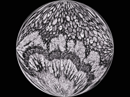
Sometimes it is nice to do something different with the images you observe through the microscope. Like in this case with a cut through the petiole of the Castanea sativa (chestnut).

Two photos were taken with the Motic BA410E. 

One with the Moticam S12, a 10X PlanApo objective, a polarization and a lambda filter. 

Another with the Moticam 285C Pro, a black and white camera. This camera is mainly intended for the recording of fluorescence images, under low light conditions. The rest is then a matter for the photo editing program.


The images are vaguely reminiscent of Mother Earth when viewed from space. You could call the images micro-worlds, so to speak. 

It is a pleasure to work with microscopy in such a way every now and then.

Friday, 24 December 2021

Not just limestone

Certain sedimentary rocks such as limestone can be impregnated with hydrocarbons and can contain 4 to 9% bitumen or "shale oil". Asphaltic or bituminous limestone is therefore a mineral impregnated with natural bitumen. This mineral is mined from underground mines or via open-pit mining. 

From asphaltic limestone, bituminous oil or asphalt can be extracted for use in the cosmetic or pharmaceutical industry. This tough and sticky product is also mixed with other raw materials to produce the well-known black pavement for our roads. In addition, it is used for waterproofing of roofs, walls, terraces, sidewalks, bridges, casings, tanks, etc. It is also a good acoustic insulator that is able to absorb the noise of machines.

Apart from the mentioned applications, the bituminous oil was once considered a universal remedy, especially good for the treatment of colds, hair loss (Hahn oil), stomach diseases and last but not least, rheumatism. By distillation of asphaltic limestone, it is still possible to obtain gas and petroleum.

On the photo taken of a thin section we see Ooids with bituminous material (containing some fossils) around it. Ooids are spherical or subspherical carbonate grains characterized by an internal concentric structure. Long time ago, the layers or coatings were formed in agitated waters as these grains were rolling around, after which they were piled in layers, under pressure, to form sediment rock.


© www.willemsmicroscope.com

Thursday, 16 December 2021

Caught and eaten by Portuguese Sundew

Portuguese Sundew or Drosophyllum lusitanicum is a shrub-like carnivorous plant that can grow up to 1.5 m in height, but is usually shorter (about 40 cm). The genus name, Drosophyllum, is derived from the Greek "drosos" (meaning dew) and "phyllon" (meaning tribe or leaf). This name is a reference to the glittering droplets on the catch stalks. 

The species name is derived from Lusitania, the ancient name for Portugal. In its native habitat, Drosophyllum is relatively rare, with plants growing in narrow coastal or maritime regions (to maximum a few tenths of kilometers from the coast) with regular morning fog during summer. 

Besides Portugal, Drosophyllum can be found in Andalusia and in northern Morocco.

Portuguese Sundew is a very interesting and curious plant for several reasons. It has two types of glands on the leaves; the huge stalked glands - as shown on the image - that attract and trap prey and the sessile, digestive glands. 

Compared with other carnivorous plants, it is one of the most successful ‘hunters’ in terms of quantity of captured prey, and all with the absolutely passive primitive adhesive traps (the stalked sticky glands). 

Production of the mucilage for trapping prey is so intense that it often drips from the leaves. Digestion of prey is very rapid, being complete within several days. Luring is accomplished by the sweet, honey-like odor of the mucilage, detectable by the human nose.


© www.willemsmicroscope.com