Thursday, 30 December 2021

Micro-worlds

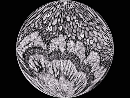
Sometimes it is nice to do something different with the images you observe through the microscope. Like in this case with a cut through the petiole of the Castanea sativa (chestnut).

Two photos were taken with the Motic BA410E. 

One with the Moticam S12, a 10X PlanApo objective, a polarization and a lambda filter. 

Another with the Moticam 285C Pro, a black and white camera. This camera is mainly intended for the recording of fluorescence images, under low light conditions. The rest is then a matter for the photo editing program.


The images are vaguely reminiscent of Mother Earth when viewed from space. You could call the images micro-worlds, so to speak. 

It is a pleasure to work with microscopy in such a way every now and then.

Friday, 24 December 2021

Not just limestone

Certain sedimentary rocks such as limestone can be impregnated with hydrocarbons and can contain 4 to 9% bitumen or "shale oil". Asphaltic or bituminous limestone is therefore a mineral impregnated with natural bitumen. This mineral is mined from underground mines or via open-pit mining. 

From asphaltic limestone, bituminous oil or asphalt can be extracted for use in the cosmetic or pharmaceutical industry. This tough and sticky product is also mixed with other raw materials to produce the well-known black pavement for our roads. In addition, it is used for waterproofing of roofs, walls, terraces, sidewalks, bridges, casings, tanks, etc. It is also a good acoustic insulator that is able to absorb the noise of machines.

Apart from the mentioned applications, the bituminous oil was once considered a universal remedy, especially good for the treatment of colds, hair loss (Hahn oil), stomach diseases and last but not least, rheumatism. By distillation of asphaltic limestone, it is still possible to obtain gas and petroleum.

On the photo taken of a thin section we see Ooids with bituminous material (containing some fossils) around it. Ooids are spherical or subspherical carbonate grains characterized by an internal concentric structure. Long time ago, the layers or coatings were formed in agitated waters as these grains were rolling around, after which they were piled in layers, under pressure, to form sediment rock.


© www.willemsmicroscope.com

Thursday, 16 December 2021

Caught and eaten by Portuguese Sundew

Portuguese Sundew or Drosophyllum lusitanicum is a shrub-like carnivorous plant that can grow up to 1.5 m in height, but is usually shorter (about 40 cm). The genus name, Drosophyllum, is derived from the Greek "drosos" (meaning dew) and "phyllon" (meaning tribe or leaf). This name is a reference to the glittering droplets on the catch stalks. 

The species name is derived from Lusitania, the ancient name for Portugal. In its native habitat, Drosophyllum is relatively rare, with plants growing in narrow coastal or maritime regions (to maximum a few tenths of kilometers from the coast) with regular morning fog during summer. 

Besides Portugal, Drosophyllum can be found in Andalusia and in northern Morocco.

Portuguese Sundew is a very interesting and curious plant for several reasons. It has two types of glands on the leaves; the huge stalked glands - as shown on the image - that attract and trap prey and the sessile, digestive glands. 

Compared with other carnivorous plants, it is one of the most successful ‘hunters’ in terms of quantity of captured prey, and all with the absolutely passive primitive adhesive traps (the stalked sticky glands). 

Production of the mucilage for trapping prey is so intense that it often drips from the leaves. Digestion of prey is very rapid, being complete within several days. Luring is accomplished by the sweet, honey-like odor of the mucilage, detectable by the human nose.


© www.willemsmicroscope.com

Wednesday, 15 December 2021

New minerals for a classic mine in Occitània: Correc d’en Llinassos, Oms, France

Oms is a beautiful village, in the ancient region of Roselló (nowadays Eastern-Pyrenees, France), in the south of historic region of Occitània. This locality includes different mining sites of scientific interest. One of these mining operations is in the Correc d'en Llinassos (torrent), near Oms (mindat.org, loc-49065).

This deposit was studied by the AFM (Association Française de Microminéralogie) in the middle of the last decade (Berbain & Favreau, 2007). In this article, numerous species were citated, the most interesting containing minerals were the Ni: annabergite, bottinoite, gersdorffite, glaucospherite, millerite, ullmannite. It is necessary to add one more that was identified for the first time worldwide: omsite, named after this locality (type locality) (Mills et al., 2017). This is a very rare hydroxyantimonite of iron, nickel and copper, member of the cualstibite group. Correc d’en Llinassos is also the type locality for hydroxyferroroméite, identified in 2017 (Mills et al., 2017a).

During a visit to the mine, with prior authorization from the owner of the farm, since access is prohibited, it was possible to collect various mineral specimens from the outer area of the mine, to later be studied. Among them, some globular aggregates of white color on siderite stood out, which after being analyzed by SEM-EDS indicated that it could be some magnesium carbonate. The Raman spectra obtained were consistent with dypingite or hydromagnesite. To close the study, a powder X-ray diffraction was carried out. Both the spectrum and the cell yielded values confirmed that it was hydromagnesite. This mineral had not been described in this mine.   

Globular white aggregates of hydromagnesite accompanied by Mg rich malachite

XRD spectrum (powder): hydromagnesite from Oms. 
Courtesy: Geociències Barcelona, Geo3BCN–CSIC, Bruker D8-A25 (Cu Kα, PSD detector)

On the other hand, it was possible to observe several globular aggregates of green colour that could correspond a visu with malachite. Reaction with HCl indicated that it was a carbonate. A copper-nickel hydroxylcarbonate from the Rosasite group has been described in this deposit, which could be identified as glaucospherite, by appearance and chemical reaction. Raman spectra also indicated that it was a similar mineral malachite. The studies were carried out using SEM-EDS and the results were very interesting since, surprisingly, most of these malachite aggregates contain a certain percentage of magnesium (Mg: Cu 1: 4-6).

Green globular aggregates of magnesium rich malachite with aragonite (white)


SEM-EDS uncoated. Malachite Mg-rich 
Cortesy: Geomar-Enginyeria del Terreny, SEM-EDS Phenom G5 XL

Its resemblance to the photographs published as glaucospherite from this deposit suggests that, in some cases, without chemical analysis it may be difficult to identify one or the other mineral. After reviewing numerous specimens, it was possible to find globular aggregates of a sky blue to bluish green color, in which magnesium and copper were found in 1: 1 proportion. It was, unsurprisingly, identified as McGinnessite.

Globular aggregates of blue-green mcguinnessite acompanied by Mg-rich malachite (green)

SEM-EDS uncoated. Mcguinnessite from Oms. 
Cortesy: Geomar-Enginyeria del Terreny, SEM-EDS Phenom G5 XL

Mineral deposits, although studies have been published on them, do not cease to amaze. Continued study will enrich the country's mineralogical heritage and contribute a grain of sand to science. Obviously, also supporting the collaboration between academic mineralogists and amateur mineralogists, mineralogists after all, is our duty and obligation. 


Acknowledgments
Thanks to Geomar-Enginyeria del Terreny for SEM-EDS studies; Dr. Jordi Ibáñez and Soledad Álvarez, Geociències Barcelona (GEO3BCN - CSIC); Dr. Tariq Jawhari, Raman departament of Centres Científics i Tecnològics de la Universitat de Barcelona (CCiTUB); Dr. Joan Carles Melgarejo (UB) to facilitate the study of the samples and to the owner of the property where the mine is located.


Bibliographical notes
- Berbain, C., Favreau, G. (2007): “Un exemple peu courant de minéralisation nickélifère: le Correc d'en Llinassos à Oms (Pyrénées-Orientales)”. Le Cahier des Micromonteurs, 95, 1, 3-24.
- Mills, S.J., Kampf, A.R., Housley, R.M., Favreau, G., Pasero, M., Biagioni, C., Merlino, S., Berbain, C., Orlandi, P. (2012): “Omsite, (Ni,Cu)2Fe3+(OH)6[Sb(OH)6], a new member of the cualstibite group from Oms, France”. Mineralogical Magazine, 76, 1347-1354.
- Mills, S.J., Christy, A.G., Rumsey, M.S., Spratt, J., Bittarello, E., Favreau, G., Ciriotti, M.E., Berbain, C. (2017a): “Hydroxyferroroméite, a new secondary weathering mineral from Oms, France”. European Journal of Mineralogy, 29, 307-314.
- Mindat.org: “Correc d'en Llinassos (Ravin d'en Llinassous), Oms, Céret, Pyrénées-Orientales, Occitanie, France”. https://www.mindat.org/loc-49065.html [on line 11/2021].

Thursday, 9 December 2021

Phacus longicauda

Phacus longicauda is a species of flattened, pointed single-celled protist microalgae belonging to the phylum Euglenozoa. It is a solitary and free-swimming algae.

Phacus longicauda has a very striking and rigid periplasm, with spiral bands, and numerous small disc-shaped chloroplasts. The cell is flat and rigid and powered by a flagellum at the front of the cell. There is a highly visible red eye spot near the flagellar base. These red eyespots are present in most Euglenoïds.

Phacus longicauda is found in freshwater habitats around the world. These habitats include ponds, pools and standing water. An organically enriched freshwater environment is essential for the development of these species.

Phacus species sometimes have a peculiar twisted shape. They are photosynthetic unicellular organisms, which means that they are able to produce their own food. Although the genus receives their nutrients primarily through photosynthesis, they are also able to feed on certain types of algae and bacteria.

Most of these organisms also have a semi-rectangular eyespot, often reddish in color, and a single flagellum, although some species have two that emerge. The flagellum is responsible for movement of the cell by rotating in the direction of travel, which allows the cell to slide and swim in the water.

At the base of the flagellum, adjacent to the eyespot, lies the paraxial swelling that functions in photoreception. It is the intricate coordinated interactions of the eyespot, paraxial swelling and some other parts of the cell that enable the cell to swim to and from the light. The ‘eyespot’ designation is therefore somewhat misplaced because it is only part of the photosensitive device and is not the photoreceptor itself, which is the paraxial swelling.

For detailed descriptions, see the standard work "Freshwater Algae of North America, Ecology and Classification".


Friday, 3 December 2021

How does it look after 4 years at about 1200 ºC?

The pictures taken with the SMZ-171 and the Moticam 10+, are showing a Haynes alloy component which has been exposed to a temperature of about 1200 oC during a period of 4 years. The component was positioned in a naphtha cracking oven (methane fuel side) of a chemical ethylene plant. On the surface of the component that is affected greatly, carburization and crystal formation can be seen. By means of ultrasonic thickness measurement it can be checked if sufficient unaffected material is left under the carburized layer, in order to see if the component is still strong enough.


Haynes 230 alloy is a nickel-chromium-tungsten-molybdenum alloy that combines excellent high-temperature strength, outstanding resistance to oxidizing environments up to 2100°F (1149°C) for prolonged exposures, premier resistance to nitriding environments, and excellent long-term thermal stability. It is readily fabricated and formed, and is castable. Other attractive features include lower thermal expansion characteristics than most high-temperature alloys, and a pronounced resistance to grain coarsening with prolonged exposure to high temperatures.

Rod material out of Haynes alloy

Exposed Haynes alloy piping distance piece

Haynes alloy 230 is composed of  Ni 57%, Cr 22%, Tu 14% and Fe < 3%

With thanks to: Gerard Janssen and Rijk Koster, mechanical engineers

© www.willemsmicroscope.com